Category:Microbiology
From WikiLectures
Pages in category "Microbiology"
The following 200 pages are in this category, out of 357 total.
(previous page) (next page)A
- A decision mechanism for the destruction of non-functional proteins
- Acinetobacter calcoaceticus
- Actinobacillus
- Actinomyces
- Actinomyces israeli
- Actinomycosis
- Acute anterior poliomyelitis
- Acute pharyngitis
- Acute viral encephalitis
- Adenoviruses
- Aeromonas and plesiomonas
- Aflatoxin
- Agents of cardiovascular infections
- Agents of Eye Infections
- Agents of Female Genital Infections
- Agents of respiratory infections
- AIDS
- AIDS diagnosis
- Allergic bronchopulmonary aspergillosis
- Amikacin
- Aminoglycosides
- Amphenicols
- Amphizoic amoebae
- Anatoxin
- Angina fever
- Anthropozoonosis
- Anti-epidemic measures in the outbreak
- Antibiotic resistance
- Antibiotic susceptibility test
- Antibiotics (neonatology)
- Antifungals
- Antisepsis
- Antistaphylococcal antibiotics and chemotherapeutics
- Archaea
- Ascariasis 1.Lf
- Aspergilloma
- Aspergillosis
- Aspergillus
- Aspergillus infections
- Aspergillus Infections
- Atlas of microbiological cultures
- Atypical mycobacteria
- Atypical mycobacterias
- Atypical pneumonia
B
- Babesiosis
- Bacillus
- Bacillus anthracis
- Bacitracin
- Bacitracin Susceptibility Test
- Bacitracin test
- Bacteria
- Bacterial gastroenteritis
- Bacterial Gastroenteritis
- Bacterial meningitis (infection)
- Bacterial meningitis (paediatrics)
- Bacterial reproduction
- Bacterial Spores
- Bacterial structure
- Bacterial toxins
- Bacterial vaginosis
- Bacterial virulence factors
- Bactericide
- Bacteriophage
- Bacteriophage therapy
- Bacteroides
- Bartonella henselae
- Beta lactam antibiotics
- Beta-lactam antibiotics
- Beta-lactamases
- Bifidobacterium
- Biochemistry of viruses
- Biofilm
- Biological weapons
- Blood agar
- Blood Agar (microbiology)
- Blood culture
- Bordetella pertussis
- Borrelia burgdorferi
- Botulism (C. Botulinum)
- Breakdown of vaccination in the Czech Republic
- Brucella
- Brucellosis
- Burkholderia
- Burkholderia cepacia
- Burkholderia mallei
- Burkholderia pseudomallei
- Burri dyeing
C
- CAMP test
- Campylobacter
- Campylobacter Enteritis
- Campylobacter enteritis 1.lf
- Candida auris
- Carbapenems
- Cat-scratch disease
- Catalase test
- Causes of Bone and Joint Infections
- Cell
- Cell nucleus
- Chickenpox
- Chocolate Agar
- Ciklopirox
- Clostridium
- Colonization
- Common cold
- Congenital syphilis
- Conjugation
- Conjugation, transformation, transduction
- Coronaviruses
- Corynebacterium
- Corynebacterium diphtheriae
- Corynebacterium jeikeum
- Covid-19
- Coxiella burnetii
- Cryptococcus neoformans
- Culture media
- Cytomegalovirus
D
- Decision-making mechanism for the destruction of non-functional proteins
- Dengue fever
- Dermatomycoses
- Dermatomycosis
- Dermatophytosis
- Development of colonization of the oral cavity by bacteria
- Development of oral bacterial colonization
- Diagnosis of Helicobacter Pylori Infection
- Diarrheal diseases (guide)
- Diarrheal diseases (signpost)
- Differential diagnosis of diarrheal diseases
- Differential diagnosis of diarrhoeal diseases
- Dimorphic fungi
- Diphtheria
- Dirofilariasis
- Disease elimination
- Disinfection and sterilization
- Disk diffusion test
- Division of vaccination in the Czech Republic
E
- E-test
- EBV
- Echinococcosis
- Echinococcus granulosus
- Echinococcus multilocularis
- Echinoviruses
- Echovirus
- Ehrlichiosis
- Elimination of infection
- Endotoxin
- Entamoeba histolytica
- Enterobacter
- Enterobiasis
- Enterococcus
- Enterohemorrhagic Escherichia coli
- ENTEROtest
- Enterotoxicosis
- Enterotoxins
- Enterovirus
- Eradication of disease
- Erysipela
- Erysipelas
- Erysipeloid
- Erysipelothrix rhusiopathiae
- Escherichia coli
G
- Gastrointestinal parasitosis
- General characteristics of parasites
- Genital herpes
- German measles
- Giardia lamblia
- Giemsa stain
- Gingivo-periodontal manifestations in viral diseases
- Glycopeptides
- Glycosylation-independent targeting
- Glycylcycline
- Glycylcyclines
- Glycylcyclins
- Gram staining
- Gram-negative anaerobic rods and cocci
- Gram-positive non-sporulating anaerobes
- Griffith's experiment
- Group A streptococcal infection
H
Media in category "Microbiology"
The following 53 files are in this category, out of 53 total.
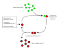
- 688px-Prion propagation.png 688 × 600; 29 KB
- Bacillus subtilis-MH agar.jpg 1,481 × 1,457; 287 KB
- Candida albicans-Sabourauduv agar.jpg 1,569 × 1,478; 277 KB
- Citrobacter freundii-DC.JPG 2,560 × 1,920; 2.16 MB
- Citrobacter freundii-krevni agar.JPG 2,560 × 1,920; 1.93 MB
- Enterococcus faecalis-krevni agar.jpg 1,333 × 1,219; 306 KB
- Enterococcus faecalis-zluc esculin.jpg 1,456 × 1,365; 266 KB
- Escherichia coli-DC.JPG 2,560 × 1,920; 2.11 MB
- Escherichia coli-Endo.JPG 2,560 × 1,920; 2.16 MB
- Escherichia coli-krevni agar.JPG 2,560 × 1,920; 1.92 MB
- Klebsiella pneumoniae-DC.JPG 2,560 × 1,920; 2.03 MB
- Klebsiella pneumoniae-Endo.JPG 2,560 × 1,920; 2.06 MB
- Klebsiella pneumoniae-krevni agar.JPG 2,560 × 1,920; 2.01 MB
- Morganella morganii-DC.JPG 2,560 × 1,920; 2.04 MB
- Morganella morganii-Endo.JPG 2,560 × 1,920; 2.04 MB
- Morganella morganii-krevni agar.JPG 2,560 × 1,920; 1.94 MB
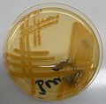
- Neisseria pharyngis-krevni agar.jpg 2,560 × 1,920; 276 KB
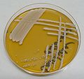
- Proteus mirabilis-DC.JPG 2,560 × 1,920; 2.05 MB
- Proteus mirabilis-Endo.JPG 2,560 × 1,920; 1.98 MB
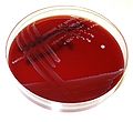
- Proteus mirabilis-krevni agar.JPG 2,560 × 1,920; 1.69 MB
- Proteus vulgaris-DC.JPG 2,560 × 1,920; 2.03 MB
- Proteus vulgaris-Endo.JPG 2,560 × 1,920; 2.09 MB
- Proteus vulgaris-krevni agar.JPG 2,560 × 1,920; 1.73 MB
- Pseudomonas aeruginosa-Endo.JPG 2,560 × 1,920; 2.03 MB
- Pseudomonas aeruginosa-krevni agar-detail.JPG 2,560 × 1,920; 1.89 MB
- Pseudomonas aeruginosa-krevni agar.JPG 2,560 × 1,920; 1.89 MB
- Pseudomonas aeruginosa-MH agar.JPG 2,560 × 1,920; 2.13 MB
- Pseudomonas fluorescens-Endo.JPG 2,560 × 1,920; 2.07 MB
- Pseudomonas fluorescens-krevni agar.JPG 2,560 × 1,920; 2.01 MB
- Pseudomonas fluorescens-MH agar.jpg 2,560 × 1,920; 2.09 MB
- Salmonella enteritidis-DC.JPG 2,560 × 1,920; 2.35 MB
- Salmonella enteritidis-Endo.JPG 2,560 × 1,920; 2.05 MB
- Salmonella enteritidis-krevni agar.JPG 2,560 × 1,920; 2.01 MB
- Shigella flexneri-DC-detail.JPG 2,560 × 1,920; 2.19 MB
- Shigella flexneri-DC.JPG 2,560 × 1,920; 2.08 MB
- Shigella flexneri-Endo.JPG 2,560 × 1,920; 2.13 MB
- Shigella flexneri-krevni agar.JPG 2,560 × 1,920; 1.97 MB
- Staphylococcus aureus-krevni agar-detail hemolyzy.jpg 2,560 × 1,920; 297 KB
- Staphylococcus aureus-krevni agar.jpg 2,560 × 1,920; 294 KB
- Staphylococcus epidermidis-krevni agar-detail.jpg 2,560 × 1,920; 2.09 MB
- Staphylococcus epidermidis-krevni agar.jpg 2,560 × 1,920; 1.96 MB
- Streptococcus agalatiae-krevni agar-hemolyza.jpg 2,560 × 1,920; 1.84 MB
- Streptococcus agalatiae-krevni agar.jpg 1,538 × 1,403; 282 KB
- Streptococcus pneumoniae M-faze-krevni agar-detail hemolyzy.jpg 2,560 × 1,920; 1.75 MB
- Streptococcus pneumoniae M-faze-krevni agar.jpg 2,560 × 1,920; 1.84 MB
- Streptococcus pneumoniae R-faze-detail hemolyzy.jpg 2,560 × 1,920; 2 MB
- Streptococcus pneumoniae R-faze-krevni agar.jpg 2,560 × 1,920; 271 KB
- Streptococcus pyogenes-krevni agar-detail hemolyzy.jpg 2,560 × 1,920; 290 KB
- Streptococcus pyogenes-krevni agar.jpg 1,387 × 1,367; 281 KB
- Symbol bacteriophage.png 50 × 50; 4 KB
- Yersinia enterocolitica-DC.JPG 2,560 × 1,920; 1.97 MB
- Yersinia enterocolitica-Endo.JPG 2,560 × 1,920; 2.12 MB
- Yersinia enterocolitica-krevni agar.JPG 2,560 × 1,920; 1.95 MB
