Culture media

Growth media are substrates that provide suitable living conditions for the growth of microorganisms. They can be used to detect and determine the properties of certain microorganisms.
Types of media
Culture media can be divided according to their state into two types: solid and liquid.
Nutrient media
They are rich in nutrients and growth factors. This includes nutrient broths.
Enriched media
They are used to cultivate more fastidious bacteria, which need special growth factors. They can be enriched with for example amino acids, vitamins, sugar etc... A typical example is blood agar enriched with a growth factor called hemin, which is necessary for the growth of Haemophilus spp. or Neisseria gonorrhoeae.
Selective media
Only certain organisms grow on selective media and are therefore used if we want to isolate a certain species. We achieve this either by adding substances that have bactericidal effects on a certain species, or by making the medium poor in certain substances (for example a certain amino acid) that are necessary for the growth of unwanted organisms. An example of a selective medium is Löwenstein-Jensen medium.
Indicator media
These growth media are used to distinguish multiple species of organisms found in one medium. This is usually achieved by a different coloration of the individual colonies, which is caused for example by different metabolism of a certain substance, the products of which then react with the indicator in the medium. An example of this is an ENTEROtest.
Selective-indicator media
They are a combination of selective and indicator media. They contain inhibitors and indicators and are therefore more specific than the previous two. Examples are Endo agar, deoxycholate citrate agar, bile esculin agar…
Transport media
They are designed to transport a clinical specimen to the laboratory. They contain a minimum amount of nutrients to avoid overgrowth and distortion of the amount of microorganism in the sample. Examples are Amies medium, Stuart medium, or Cary-Blair medium.
Specific examples of media
Nutrient broth
- a universal liquid medium
- microbial growth is indicated by turbidity in an originally clear medium (facultative anaerobes), surface turbidity (aerobes), or a sediment (anaerobes)
- by adding agar (seaweed polysaccharide) it is made into solid media known as agar
Nutrient media
- they contain essential nutrients for the growth of bacteria
- they can be liquid (broth) or solidified with agar
- they are the base for enriched media
Blood agar
- a dark red medium, made of a nutrient medium enriched with blood (mostly sheep or horse) and a natural polysaccharide agar
- both G+ and G− bacteria grow on it
- it is suitable for most species
- hemolytic bacteria that produce hemolysin make the agar lighter – which is used in the CAMP test to differentiate Streptococcus agalactiae and Streptococcus pyogenes
Chocolate agar
- it is made by adding blood to an agar base at a temperature of 85°C[1]
- the denaturation of blood by the heat is responsible for the brown (chocolate) color
Endo agar
- it is light pink and turns more pink when exposed to light
- G+ bacteria do not grow on it
- distinguishes lactose-positive bacteria from lactose-negative bacteria. Lactose-positive bacteria metabolize lactose – they create aldehydes, which react with fuchsine in the Endo medium and create Schiff bases, turning the area around the colonies dark pink.
- Lactose-negative bacterial colonies do not induce this color change (the color remains the same).
MacConkey agar
- Similarly to the Endo agar, it distinguishes lactose-positive bacteria from lactose-negative bacteria. Lactose-positive bacteria turn the medium pink or red, and lactose-negative bacteria do not.
Deoxycholate citrate agar (DC agar)
- G+ bacteria do not grow on it
- G- bacteria that ferment lactose – pink-red color
- lactose-negative – white (for example some intestinal bacteria)
- contains Na2S2O3 (sodium thiosulphate), which together with hydrogen sulfide, that is produced by certain bacteria species (Salmonella, Citrobacter, Proteus), creates Fe sulfides – black tips on the colonies
- suppresses the swarming growth of Proteus
Bile Esculin agar (BE agar)
- used to diagnose G+ Enterococcus faecalis (large intestine) → urinary tract infections
Clauberg agar
- among other ingredients, it contains tellurite, which inhibits the growth of most bacteria except for Corynebacterium, which reduce tellurite to tellurium, and that is subsequently reflected in the color of the agar (gray, brown or black).
Tinsdale Agar
- Similar to the Clauberg agar, it is used to identify Corynebacterium spp.
- a gray-black halo is formed around the colonies as a product of the reaction of tellurium with sulfane (formed from cystine)
Löwenstein-Jensen agar
- used to cultivate mycobacteria
Sabouraud agar
- it is a light yellow medium, which is too poor in nutrients for bacteria, but yeasts and fungi grow on it
Mueller–Hinton agar (MH agar)
- it is used to determine antibiotic susceptibility (disk diffusion method)
- there is also a kind enriched with blood
Gallery of culture media
-
 Bacterial smear on nutrient agar Escherichia coli
 -
 Blood agar with Enterococcus faecalis
 -
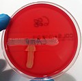 CAMP test on blood agar
 -
 Chocolate agar
 -
 Endo agar
 -
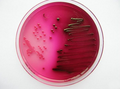 Escherichia coli on Endo agar
 -
 Escherichia coli on MacConkey agar
 -
Lactose-positive (pink, on the left) and lactose negative (light orange – the same colour as the medium, on the right) colonies on MacConkey agar
-
Salmonella sp. on deoxycholate citrate agar
-
 Enterococcus faecalis on bile-esculin agar
 -
Löwenstein–Jensen agar
-
Löwenstein-Jensen agar with Mycobacterium kansasii
-
 Sabourad agar with Candida albicans
 -
MH agar with MRSA


References
Related articles
Sources
- RYŠKOVÁ, Olga, et al. Návody k praktickým cvičením z lékařské mikrobiologie. 1. edition. Praha : Karolinum, 1997. ISBN 80-7184-307-5.
- JULÁK, Jaroslav. Praktická cvičení a semináře z lékařské mikrobiologie. 2. edition. Praha : Karolinum, 2009. 113 pp. ISBN 978-80-246-1141-9.
Citations
- ↑ JULÁK, Jaroslav. Praktická cvičení a semináře z lékařské mikrobiologie. 2. edition. Praha : Karolinum, 2009. 113 pp. ISBN 978-80-246-1141-9.


