Necrosis

Necrosis is intravital tissue death'. It includes the whole spectrum of morphological changes that take place after the death of the cell. Necrotic changes take several hours to develop and are therefore not macroscopically apparent immediately after cell death.
Infarction (ischemic necrosis):
Infarction (ischemic necrosis) 2:
It is mostly a pathological process, but it can also be part of physiological processes (e.g. an alternative way of eliminating tumor cells that inactivate apoptosis).

| Criterion | Necrosis | Apoptosis' |
| Cell Size | ↑↑↑ | ↓↓↓ |
| Core | Basophilia disappears, decay | Chromatin condensation |
| Plasma Membrane | Rupture | Formation of apoptotic bodies |
| Organelles | Zoom out + swell | Aggregation + lysis |
| Inflammatory reaction | Yes | No |
| Genetics | (No) | Yes |
| Occurrence | Anywhere | Rapidly dividing tissues of the embryo, involution |
Most common causes[edit | edit source]
- Action by physical factors' (mechanical damage, burns, frostbite, electric current, UV radiation, radiation);
- action by chemicals' (acids, hydroxides, substances in toxic concentration, drugs, poisons, toxins, asbestos, free radicals);
- ischemia, hypoxia (anemia, CO poisoning, lung damage);
- immune disorders (autoimmune disease, allergy);
- infectious agent (bacteria, viruses).
Manifestations of necrosis[edit | edit source]
Microscopic Manifestations[edit | edit source]
Breakdown of nuclear DNA causes loss of basophilia (karyolysis). Chromatin can accumulate on the inside of the nuclear membrane (mural hyperchromatosis). Subsequently, the nucleus breaks up into small fragments (karyorhexe). Another way of death is cell shriveling with increased basophilia of the nucleus (pyknosis). In both cases, the core disappears within one or two days. The cytoplasm becomes more eosinophilic. Organelles (mainly mitochondria) swell. There is vacuolization and subsequent disruption of the membranes of the organelles and the cell itself. Disruption of the membrane of lysosomes causes the release of their enzymes into the environment, which results in self-digestion of the cell (autolysis).
Proteins from dying cells are released into the environment and can be recognized by the immune system as foreign. inflammatory reaction occurs. The presence of this tissue reaction distinguishes necrosis from postmortem autolysis. polymorphonuclear cells and macrophages emerge from the blood vessels around the cell (chemotaxis - attracted by inflammatory mediators). These release enzymes that enter the necrotic cells and cause their digestion (heterolysis). Necrotic cells can be removed by phagocytosis and replaced by new tissue.
Biochemical manifestations[edit | edit source]
The most important biochemical manifestation of necrosis is a decrease in tissue pH. There is an influx of Ca2+, which activates enzymes and thus contributes to autolysis. Enzymes released from lysosomes and monocytes cause subsequent changes in the tissue by denaturing proteins.
We use the released enzymes as "serum markers" of organ damage. These include, for example: transaminases, lactate dehydrogenase, creatine kinase, alkaline phosphatase and acid phosphatase.
Clinical manifestations[edit | edit source]
Necrosis is usually accompanied by an inflammatory reaction. Clinical manifestations include increased sedimentation and temperatures. There is leukocytosis and the formation of edema, or ulceration.
Distribution of necrosis[edit | edit source]
Simple necrosis[edit | edit source]
It affects the skin and muscles when the supply arteries are blocked. Macroscopically, it is unremarkable. The muscles are light, brownish-red and tear easily. Microscopically, there is a loss of staining of the nuclei and an increase in eosinophilia of the cytoplasm.
Colicative necrosis (malacia)[edit | edit source]
Total enzymatic decomposition of the tissue, manifested by liquefaction (colliquation). The condition is massive autolysis and heterolysis. The components of a given tissue must be easily degradable. It is most often found in ``protein-poor tissues that contain more water (white matter of the brain). The consistency of the tissue is already soft from the start. The final stage of enzymatic digestion is a pseudocyst - a pathological cavity without its own lining (bounded by tissue in which necrosis has occurred). It occurs, for example, in the brain during ``encephalomalacia or during purulent inflammatory processes (bacterial infection, mycosis), when a cavity filled with pus (abscessu) occurs.
- Liquifactive necrosis kolikvacni nekroza 20x.jpg
Colicative necrosis - Liquifactive necrosis kolikvacni nekroza 4x.jpg
Colicative necrosis 
Pseudocyst 
Encephalomalacia 
Pseudocyst Pancreas
_(14782324284).jpg)
_(14798066953).jpg)

Coagulation necrosis[edit | edit source]
Typical for hypoxic death of cells of parenchymatous organs rich in proteins (liver, kidney, myocardium, spleen). It is most common during heart attacks. It arises as a result of protein denaturation of the affected tissue. Macroscopically, it resembles boiled (coagulated) meat. The tissue is stiff and yellowish. tissue fluid penetrates the damaged area, and thus edema occurs. It tends to be wedge-shaped (typically in the kidneys). A ``hemorrhagic rim (stretched, congested capillaries) is formed around the focus of necrosis.
- Soubor:Acute myocardial infarction with coagulative necrosis (4).JPG
Heart-attack - Soubor:HE (myocardium) HE (myokard) 20x.jpg
Heart-attack - Soubor:infarction of myocardium infarkt myokardu 4x.jpg
Heart-attack 
Heart-attack 
Kidney infarction


Hemorrhagic necrosis, caseification necrosis and Zenker's muscle necrosis are special cases of coagulation necrosis.
Hemorrhagic necrosis[edit | edit source]
It is caused by massive bleeding of necrotic tissue (when tissue fluid penetrates the affected area, erythrocytes and plasma from the circulation also pass through). It can arise as a result of blood flow stasis during vein obstruction (venous thrombosis). Another cause may be the creation of negative pressure in the tissue area (obstruction of the terminal artery) and subsequent reflux from the capillary and venous low-pressure areas into the surrounding tissue. Macroscopically, it manifests as a red-violet color of the tissue (if the cause of necrosis is ischemia, it has the appearance of a so-called red heart attack - lungs, spleen, adrenal glands). It occurs, for example, in ``red encephalomalacia, which arises from thrombosis of intracranial vessels or, for example, in the cortex of the brain, where there is a possibility of capillary reflux). More rarely, also with massive toxic necrosis of capillaries in hemorrhagic-necrotic inflammations (plague).
- Soubor:hemorrhagic necrosis colon hemoragicka infarzace streva 20x.jpg
Hemorrhagic intestinal infarction - Soubor:hemorrhagic necrosis colon hemoragicka infarzace streva 4x.jpg
Hemorrhagic intestinal infarction 
Hemorrhagic pulmonary infarction - Soubor:hemorrhagic necrosis lung hemoragicka nekroza plice 4x.jpg
Hemorrhagic pulmonary infarction - Soubor:Kidney – acute cortical necrosis.jpg
Acute cortical necrosis of the kidney

Caseification (caseous, powdery) necrosis[edit | edit source]
It arises during specific chronic inflammation' (immunopathological reaction of type IV). An example can be tuberculosis, some mycoses and syphilis. Macroscopically, it is granular, yellowish (caused by lipids that are released from the tissue), resembles cheese (crab). The affected area is bordered by specific granulation tissue (epithelioid and Langhans cells arising from histiocytes). Typical for this type of necrosis is eosinophilic staining of the cytoplasm with basophilic dust (fragments of the nuclei of disintegrating cells). If the infection clears, caseous necrosis calcifies and is easily detected by X-ray examination.
- Soubor:caseous necrosis lung kaseozni nekroza plice 20x.jpg
Caseous necrosis - Soubor:caseous necrosis lung kaseozni nekroza plice 4x.jpg
Caseous necrosis 
Tuberculosis of the lungs CT 
Caseous necrosis of the lungs 
Caseous necrosis of the lungs


_focus_(6596011395).jpg)
Zenker's wax necrosis[edit | edit source]
Coagulation necrosis of skeletal muscles in some infectious diseases (eg abdominal muscles in Typhoid typhus, influenza, or tetanus). Macroscopy is mostly unremarkable, the muscles look like they have been boiled, at other times they are clearly bloodied. They crack at the site of necrosis. The cross-striation of the muscle fibers is lost, and they break up lumpily (eosinophilic clumps of sarcoplasm are evident).
Fat tissue necrosis[edit | edit source]
Circumferential breakdown of fatty tissue caused by release of pancreatic enzymes (into the affected area of the pancreas, into the peritoneal cavity). It can occur as post-traumatic necrosis or as Balser's necrosis. Balser's necrosis are caused by the action of released proteolytic and lipolytic enzymes of the necrotic pancreas (acute pancreatic necrosis) on fat tissue.
During adipocyte necrosis, triacylglycerols are released and hydrolyzed. This results in the release of fatty acids, which crystallize and turn into calcium salts. It creates chalky white deposits that are visible to the naked eye.

Fat tissue necrosis 
Fat tissue necrosis


Fibrinoid necrosis[edit | edit source]
A regressive change in connective tissue' (connective tissue, vessel walls) that is immunely conditioned (hypersensitivity II. and Type III). It most often occurs in rheumatoid arthritis (affects the heart and joints), polyarteritis nodosa and collagenoses - systemic lupus erythematosus' ', scleroderma, dermatomyositis. Activation of complement and chemotaxis occurs. Here, the leukocytes break down and release proteolytic and hydrolytic enzymes that break down the collagen. Macroscopically, no significant changes are visible. Vascular wall bulges and rheumatic nodules on the heart valves may be present. Microscopically, the presence of fibrinoid, which is an amorphous mass that stains like fibrin, is significant. It is caused by the loss of the fibrous arrangement of collagen and the penetration of the tissue by fibrinous exudate. Fibrinoid stains the same as fibrin (hence the name fibrinoid necrosis).

Fibrinoid necrosis tepny
.jpg)
Bone tissue necrosis[edit | edit source]
It affects bone tissue and marrow structures (ligament, fat tissue). It arises as a result of trauma, ischemia, radiation, use of corticoids. The causes may not always be known (idiopathic avascular necrosis in children). Pyknosis of osteocyte nuclei occurs in the center of necrosis. On the periphery, there is karyolysis and the presence of empty lacunae (oncosis). At the same time, tissue and fat necrosis of the bone marrow (conspicuous edema) may occur. It is mostly hemorrhagic in nature.

Necrosis of bone tissue 
Necrosis of bone tissue


Another Fate of Necrosis[edit | edit source]
The tissue that succumbs to necrosis is destroyed by neutrophil enzymes by heterolysis. Subsequently, there is granulation of the tissue (ingrowth of hair follicles and fibroblasts). The tissue is bordered (demarcation) by polymorphonuclear cells (they penetrate only where pO2 is non-zero) and absorbed by macrophages. Further development depends on the size of the bearing.
Small bearing[edit | edit source]
It depends on the tissue in which the necrosis has occurred. In the liver, ligament or bone, regeneration takes place, which means replacement of the affected tissue with morphologically and functionally equivalent tissue. In the ``myocardium or the ``CNS there is a ``scar reparation (loss of blood vessels, fibroblasts turning into fibrocytes, tissue formation). In the ``brain and in the ``pancreas there is colic and the formation of a ``pseudocyst.
Large bearing[edit | edit source]
There will be a fibrous border (encapsulation of necrotic tissue) which may thicken and calcify (typically caseous masses in tuberculous granulomas).
Another mechanism is sequestration, in which necrotic tissue is separated from vital tissue by heterolysis. The sequestrum may float in a pseudocyst (pancreas) or, if it is on the surface (skin, mucosa), it may excoriate to form an ulcer that heals with a scar.
Secondary changes of necrosis[edit | edit source]
They arise as a result of various influences on necrotic tissue. We call secondary altered necrosis gangrene.
Clinical example[edit | edit source]
Acute myocardial infarction (heart attack): Occlusion of a coronary artery causes ischemia of the myocardium leading to coagulative necrosis of cardiac muscle. Clinically this may present with severe chest pain, diaphoresis, and elevated cardiac troponins; necrotic tissue later triggers inflammation and is replaced by scar.
Secondary modifications include:
- Desiccation resulting in dry gangrene (mummification).
- Modification of infections, when putrefactive bacteria get into the necrotic focus, which causes the formation of 'moist gangrene.
- Infections with clostridia that cause gassy gorse' (gangraena emphysematosa).

Necrosis of bone tissue 
Dry gangrene 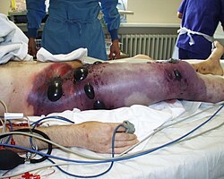
Gas gangrene


Links[edit | edit source]
Related Articles[edit | edit source]
External links[edit | edit source]
- PASTOR, Jan. Langenbeck's medical web page [ online ]. © 2006. [ cit. 2009-09-01 ]. <https://langenbeck.webs.com/>
Literature used[edit | edit source]
- POVÝŠIL, Ctibor and Ivo ŠTEINER, et al. General pathology. 1st edition. Prague: Galén, 2011. 290 p. ISBN 978-80-7262-773-8.
References[edit | edit source]
- KUMAR, Vinay; ABBAS, Abul K.; ASTER, Jon C. Robbins & Cotran Pathologic Basis of Disease. 10th edition. Philadelphia: Elsevier, 2021. ISBN 978-0-323-60992-0. (Chapter: Cell injury and cell death; ischemic necrosis/infarction).