Edema
Edema (lat. oedēma, -tis, n. , honorifically often edema) is the accumulation of fluid in the intercellular space. From the intercellular space, it can also get into other spaces – e.g. into body cavities or alveoli. It is a local blood circulation disorder, but it can also have a cause outside the cardiovascular system. As a result of the swelling, the exchange of water and nutrients between the blood and the cells is blocked, as a result of which the metabolic conditions change.
Edema:
Clinical picture[edit | edit source]
Swelling affects organs with a lot of loose connective tissue, little parenchymatous organs. The edematous organ is soaked, swollen, pasty , and when pressed, a depression forms in it (pitting edema). The skin over the swollen area is shiny, tense and pale (with the exception of inflammatory swelling), and folds after the swelling subsides.
Microscopy[edit | edit source]
The cells are distant and enlarged (swelling of cells and interstitium), between the bundles of the ligament there are slits filled with edema fluid.
Causes of swelling[edit | edit source]
The causes of swelling are pathological conditions in which the leakage of fluid into the extravascular space prevails over its reabsorption into the blood. They can be the result of a change in the hydrostatic gradient, a change in the osmotic gradient (incl. blockage of lymphatic drainage), changes in the permeability of the capillary wall or as a combination of these three mechanisms.
Division and differential diagnosis[edit | edit source]
The essential clinical distinction is at[1]: :
- generalized swelling and
- local swelling.
Pathophysiologically, swelling is divided into:
- venostatic – generalized in heart failure and local in venous swelling (phlebothrombosis, venous insufficiency),
- hypalbuminotic – only generalized, in liver or renal edema,
- lymphostatic - only local,
- toxic - only generalized in liver edema,
- inflammatory - only localized in inflammation, sarcoidosis - and
- angioneurotic – only generalized in allergies, angioedema and C1 protein defect.
Generalized swelling[edit | edit source]
Generalized edema can be venostatic, hypalbuminotic, toxic and angioneurotic. Clinically, cardiac, renal, hepatic, hypalbuminemic, allergic, preeclampsia, and false swelling of the myxedema type are distinguished.[1].
In generalized edema, the renin - angiotensin - aldosterone axis is always activated due to the reduced volume of circulating fluid.[1].
Cardiac edema[edit | edit source]
Etiopathogenetically, it is venostatic edema. The cause is the failure of the heart as a pump, the result of which is an increase in the hydrostatic pressure at the venous end of the capillary. This will increase water filtration from the capillary into the tissue and decrease reabsorption. Fluid escapes into the interstitium until the hydrostatic pressures equalize. By reducing the volume of the intravascular bed, the RAA loop is activated via baroreceptors, which causes water retention.
- Clinical picture
In right-sided heart failure, the basic manifestation is the formation of symmetrical perimalleolar swelling (in standing or sitting patients, in lying patients the back area and genitals are swollen), which spreads upwards. Furthermore, increased filling of jugular veins with pulsation, hepatojugular reflux, hepatomegaly, splenomegaly and ascites.[1].
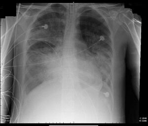
In left-sided heart failure, it is pulmonary edema (first interstitial, if the hydrostatic pressure of the fluid exceeds the pressure of the alveolar air, alveolar edema occurs).
- Diagnostics
The basis is ECHO. Changes are also visible on X-ray (pulmonary edema) or ECG.[1].
Liver swelling[edit | edit source]
Hepatic edema combines two mechanisms: toxic and hypalbuminemic. The basis is blood stasis in the portal system, which is penetrated by gram-negative intestinal bacteria, which are normally eliminated in the liver. Stasis opens portocaval anastomoses, through which Gram-negative bacteria enter the systemic circulation. This endotoxemia causes an increased production of nitric oxide , which has a vasodilating effect in the periphery. The result is swelling, a decrease in circulating blood volume and activation of the renin-angiotensin-aldosterone system.
The damaged synthetic function of the liver also causes hypalbuminemia and a drop in oncotic pressure (see below).
- Causes
The most common cause is liver cirrhosis as a result of ethylism.[1].
- Diagnostics
History (ethylism, bleeding from esophageal varices, ascites puncture), physical examination (hepatomegaly, ascites), sonography of the abdomen (tumors). Biochemically, liver function, albumin (prealbumin).
Renal edema[edit | edit source]
Renal edema can be caused by two mechanisms: sodium and water retention (oliguria / anuria) and hypalbuminemia (proteinuria, see below).
- Diagnostics
Sonography of the kidneys is important in diagnosis, in addition to biochemical examination.[1].
Hypoalbuminotic edema[edit | edit source]
Pathophysiologically, it is a lack of plasma proteins (especially albumins), the consequence of which is a drop in blood oncotic pressure. Thus, the reabsorption of water from the interstitium into the capillaries decreases, the liquid penetrating the interstitium increases its hydrostatic counterpressure, and once the balance is established, swelling stops. Loss of intravascular fluid activates the RAA system, which, by affecting renal function, causes water retention.
- Diagnostics
History and physical examination. The diagnosis is primarily based on evidence of hypalbuminemia. There is also an examination of the synthetic function of the liver, urine biochemistry, and ultrasound of the abdomen.
- Causes
It is either an insufficient supply of protein through food or a loss of blood proteins[1]. Insufficient supply is common in eating disorders, dysphagia, malabsorptive conditions or cancer. Increased protein loss can take place in the urine in the form of proteinuria, in the stool in exudative enteropathy or in effusions (ascites, fluidothorax).
Angioneurotic edema[edit | edit source]
It is an increase in capillary permeability due to the effect of various toxins (insect stings...), in sepsis, shock, inhalation of irritating substances in the lungs, etc.
We distinguish three types[1].:
- edema on the basis of IgE-mediated reactions in allergies,
- angioedema ,
- rare edema on the basis of hereditary or acquired defect of C1 protein inhibitor.
Edema in preeclampsia[edit | edit source]
It is the release of angiogenic factors from the placenta as a result of placental hypoxia. The result is systemic endothelial dysfunction. Swelling is aggravated by proteinuria.[1].
Drug swelling[edit | edit source]
It is a side effect of the medication. E.g. swellings are common with calcium channel blockers[1], corticoids, hormonal contraception.
Cyclic swellings[edit | edit source]
Some women experience premenstrual swelling, the etiology of which is unknown.[1].
False swelling of the myxedema type[edit | edit source]
Myxedema is a swelling resembling a condition in very advanced hypothyroidism. It is not the accumulation of fluid itself in the interstitium, but also the accumulation of mucopolysaccharide.[1].
- Clinical picture
Unlike true edema, myxedema is hard. The clinical picture is complemented by fatigue, somnolence and a slow, husky voice. Bradycardia is severe in hypothyroidism. It is necessary to pay attention to cardiac arrest.
- Therapy
Thyroid hormone replacement.
Local swelling[edit | edit source]
Lymphostatic edema (lymphedema)[edit | edit source]
When the lymphatic drainage of the interstitium is impaired, proteins accumulate in it and the osmotic pressure of the interstitium increases. Lymphedema is a localized, stiff, asymmetric swelling (on the limb it leads to elephantiasis). The cause is blockage of lymph nodes and blood vessels.
It can be primary - congenital deficit development of lymphatic vessels (hypoplasia of lymphatic capillaries and precollectors). Hereditary lymphostatic edemas are rare, in which the clinical picture is enhanced by yellow nails[1]. Primary lymphedema is usually ascending (periphery is first affected and later spreads proximally).
Secondary lymphedema occurs secondary to:
- streptococcal infections - erysipelas,
- parasites - filariasis (hairworms – Filaria),
- mechanical disruption of lymphatic vessels during an accident or surgery (e.g. removal or irradiation of lymph nodes as part of anti-tumor treatment),
- tumor (most often cancer metastases in the lymph nodes),
- extirpation of the great saphenous vein for bypass operations,
- inflammation.
Secondary lymphedema is usually descending.
Therapy[edit | edit source]
The basis of treatment is physical therapy (lymphatic drainage), compression bandages and stockings, skin care, regimen measures (regular care of the limb, avoiding even minor trauma, staying in a hot environment, intensive physical work and sports).
Inflammatory edema[edit | edit source]
Due to the influence of inflammatory mediators, capillary permeability for high-molecular substances (exudation) increases.
- Causes
The most common cause is non-specific inflammation. Swelling is one of the classic signs of inflammation (tumor). A rare transverse swelling is, for example, sarcoidosis, in which the swelling can appear as erythema nodosum.
Venous swelling[edit | edit source]
It is swelling due to blockage of venous flow or venous insufficiency.
- Clinical picture
Inflammatory swelling is distinguished from other (pale) swellings by its livid color. The picture is fundamentally different if the swelling is acute or chronic[1]. Acute swelling arising mostly on the base can reach the image of phlebothrombosis phlegmasia cerulea dolens. The limb is then swollen, cyanotic and very painful. Chronic swelling arises on the basis of chronic venous insufficiency . The picture of swelling is complemented by the presence of phlebectasies (corona phlebectatica) and varices, pigmentation disorders (hemosiderin pigmentation), stasis dermatitis, white atrophy, lipodermatosclerosis or leg ulcers.
- Causes
Phlebothrombosis in acute cases , venous insufficiency in chronic cases. Beware of impending pulmonary embolism in phlebothrombosis.
Lipedema[edit | edit source]
Lipedema is hyperplasia of subcutaneous fat tissue. It occurs in women, often with a family predisposition.
Orthostatic swelling[edit | edit source]
They arise when standing and sitting for a long time, when the muscle pump is not working.
Edema fluid[edit | edit source]
- protein-poor (transudate) – soft swellings – venostatic and hypalbuminotic edema,
- rich in proteins (exudate) - stiff swellings - lymphostatic and inflammatory edema.
Light's criteria are used to differentiate between transudate and exudate.[1].
Special cases and terms[edit | edit source]
Pulmonary edema
- occurs with left-sided heart failure (clogged blood in the lungs) – fluid penetrates into the alveoli,
- clinically - coughing up pink foamy sputum, later the sputum is rusty,
- during long-term stasis, erythrocytes disintegrate and hemoglobin is converted into hemosiderin, which is phagocytosed by alveolar macrophages (siderophages), which also enter the edema fluid (hence the rusty color of the sputum), interstitial tissue multiplies,
- macroscopically: the lung is enlarged, stiff, seeped, foamy clear fluid flows from it on the cut, in longer-term conditions the lung is rust-colored and stiff (rusty induration of the lung).
- macroscopically: the brain is enlarged, heavier, narrowed sulci, flattened gyri due to pressure on the calve, narrowed ventricles (in contrast to brain atrophy, where the ventricles and sulci are enlarged and the gyri are narrowed, an atrophic brain has less weight),
- pressure cones are created (conus occipitalis, temporalis, interhaemisphericus, frontalis),
- causes death.
- Hydrops
- generalized edema with the presence of edema fluid (transudate) in preformed cavities (serous cavities, joints).
- hydrothorax (in the pleural cavities),
- hydropericardium (in the pericardial sac),
- hydroperitonaeum = ascites (in the peritoneal cavity),
- hydrocele (in cavum serosum scroti).
- Hydrops fetus universalis
- e.g. with Rh incompatibility (mother Rh-, father Rh+, child Rh+).
- Anasarka
- connective tissue edema ("generalized tissue oozing").
- Maceration
- softening of the tissue by the action of water (mainly postmortem in fetuses that died in utero).
Links[edit | edit source]
Related links[edit | edit source]
External links[edit | edit source]
- Swelling (Czech Wikipedia)
- Edema (English Wikipedia)
- Edema in venous insufficiency
References[edit | edit source]
Zdroj[edit | edit source]
- PASTOR, Jan. Langenbeck's medical web page [online]. [cit. 2009]. <http://langenbeck.webs.com>.
- ČEŠKA, Richard, ŠTULC, Tomáš, Vladimír TESAŘ a Milan LUKÁŠ, et al. Interna. 3. edition. Praha : Stanislav Juhaňák - Triton, 2020. 964 pp. ISBN 978-80-7553-780-5.


