Ligament

Ligament is the most widespread type of connective tissue in the human body. It contains cells surrounded by a large amount of intercellular material, which has a gel-like consistency and is reinforced by fibers. The main function of the ligament is to connect epithelial tissue with other tissues (e.g. muscle).
Tissue cells[edit | edit source]
Cells fixed
We include cells that are permanently settled in the ligament. Their precursor is a classical mesenchymal cell. Fixed cells include: fibroblast, fibrocyte, myofibroblast, reticular cell, melanocyte, and adipocyte.
Stray cells
Cells that entered the tissue only secondarily. The origin is primarily blood cells. Stray cells include: heparinocytes, macrophages and plasma cells.
Fixed cells[edit | edit source]
Fibroblast[edit | edit source]
It is a basic cell that is mitotically and synthetically active. It is capable of creating all types of fibrous and amorphous intercellular matter (collagen, elastin, GAG, multiadhesive glycoproteins, etc.) It is among the most widespread cell types in fibrous tissue. Like other fixed cells, it arises from mesenchyme. Elongated, star-shaped with many irregular projections. Roughly in the middle of the cell lies a large ovoid nucleus containing pale euchromatin with prominent basophilic nucleoli. Inside the cell there is a massively developed GER and the Golgi complex. The cytoplasm of the fibroblast is basophilic. Once the fibroblast is surrounded by the newly synthesized extracellular matrix, it becomes a fibrocyte.
Fibrocyte[edit | edit source]
A resting form of a fiber cell that arises from a fibroblast surrounded by an extracellular matrix. It loses its synthetic and mitotic activity and becomes the structural support of the ligament. The cell is spindle-shaped and smaller than a fibroblast. It contains a small longitudinal nucleus that is smaller and darker than that of a fibroblast and its cytoplasm is acidophilic (less GER than that of a fibroblast). Fibrocytes can retrograde into fibroblasts (e.g. during wound healing).
Myofibroblast[edit | edit source]
It closely resembles a fibroblast, but has some properties in common with smooth muscle. It produces α-smooth muscle actin, which is anchored in dens bodies along the cell edge. Actin anchored in this way runs across the cell and enables its contraction. It differs from a muscle cell in that it does not have a basement membrane.
Reticular cell[edit | edit source]
A variant of fibroblast specialized in the formation of reticular fibers, which arise from type III collagen precursors. It arises from mesenchyme. It is star-shaped with very long projections that touch each other to form a three-dimensional network.The projections of the reticular cells are in contact with the reticular fibers. This connection creates a support network for wandering cells (macrophages, lymphocytes, hematopoietic cells). Reticular cells are found mainly in the peripheral lymphatic organs, except for the thymus, in which it forms supporting tissue.

Melanocyte[edit | edit source]
An irregularly shaped pigment cell that arises from neuroectoderm (not mesenchyme!!). Inside the cell, the brown pigment melanin is synthesized and collected. The cells contained in the ligament are star-shaped. Some melanocytes communicate with epithelial cells by means of projections (pigment release). Such cells form protrusions only in the part that communicates with the epithelium. On the opposite side of the cell lies the nucleus. The cell also contains mitochondria, GER and SER.
Fat cell (adipocyte)[edit | edit source]
It is found either singly or in small groups in the ligament. The precursor is an adipoblast (from a mesenchymal precursor cell). It belongs to conspicuously large cells in the connective tissue. They pick up neutral lipids from the bloodstream, which they collect (in the form of triglycerides), enable their metabolism and release them back. They are separated from the surrounding structures by an external lamina, which resembles the basal lamina of the epithelium. The adipocyte is the basic component of adipose tissue and can make up up to 30% of body weight. On normal histological preparations, adipocytes appear as empty cells (fats were extracted during preparation of the preparation).
According to the form of fat storage inside the cell, we divide adipocytes into two groups - multivacuolar and univacuolar.
Univacuolar adipocyte[edit | edit source]
A spherical cell that is mostly made up of one large fat droplet. The cytoplasm together with the heavily flattened nucleus and other cell organelles is pushed to the periphery of the cell. The GER and Golgi complex are poorly developed, while the SER is massively developed. It also contains pinocytotic vesicles and microtubules. Univacuolar adipocytes form the main component of white adipose tissue.
Multivacuolar adipocyte[edit | edit source]
A cell containing a large number of tiny droplets of fat. The spherical nucleus is located irregularly, rather in the center of the cell. There is a large number of mitochondria in the cytoplasm. It forms the main component of brown adipose tissue. They convert chemical energy into heat (the thermoregulatory function of brown adipose tissue in newborns).

Fibroblasts 
Fibrocytes 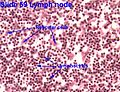
Reticular cells 
Melanocyte 
Adipocytes


Stray cells[edit | edit source]
They are formed in the red bone marrow. Their precursor cell is a hemocytoblast. They reach the ligament through the bloodstream.

Macrophage (histiocyte)[edit | edit source]
It differentiates from blood monocytes, which mature into macrophages after penetrating the tissue. The shape of the cell is variable, forming a large number of projections. Inside the cell there is a kidney-shaped nucleus, a large number of lysosomes and elements of the cytoskeleton. GER and Golgi complex are massively developed. Histiocytes move amoeboidally. It belongs to the monocytomacrophage system, therefore it has the ability of phagocytosis.
Macrophages are part of the immune system. They are able to phagocytose damaged or dead cells, possibly foreign cells, particles and microorganisms. They create highly reactive particles with the ability to destroy absorbed organisms. They release anti-inflammatory mediators. They occur mainly in the sparse connective tissue along the capillaries.
Plasma cell (plasmocyte)[edit | edit source]
Plasma cells are ovoid to spherical cells. Their characteristic appearance is due to the eccentrically placed nucleus, which is shifted to one pole of the cell. In the nucleus, dark heterochromatin alternates with light euchromatin, creating a bow-shaped arrangement. They contain a large amount of cytoplasm, which is filled with rich GER (intensive proteosynthesis). Near the nucleus, there is a bright part of the cytoplasm, which corresponds to the location of the Golgi apparatus. They arise from B-lymphocytes and are responsible for humoral immunity (production of antibodies). There are very few of them in the connective tissue, they are mostly contained in places where bacteria and foreign substances easily penetrate into the body.
Mast cell (heparinocyte)[edit | edit source]
A relatively large ovoid-shaped cell, also called a mast cell. Their cytoplasm is filled with large basophilic granules. Basophilic granules are filled primarily with heparin and histamine, which show metachromasia (when stained, the resulting shade of color is different from the dye used), stained with toluidine blue. They occur in the thin collagenous tissue of the mucous membranes of the organs of the digestive and respiratory systems. They are important for the correct course of the inflammatory reaction. It mediates the body's allergic reaction to an antigen. The mast cell membrane has an affinity for IgE. When antigen binds to IgE, basophilic granules are washed out (degranulation) into the intercellular space. This phenomenon can also occur when irritated by chemical or physical influences.

Macrophages 
Plazmocyte


Intercellular mass of ligament[edit | edit source]
Ligament fibers[edit | edit source]
Collagen fibers[edit | edit source]
Macroscopically, they have a white to silver color. They are not branched, have high tensile strength, but are not very stretchy. They contain amino acids: glycine, proline, hydroxyproline. Since glycine is the smallest amino acid, it enables the spatial arrangement of collagen into a triple helix.
We distinguish several types of collagen
- Collagen I – (most common type, 90%) – Made up of fibrils that form bundles of fibers. The individual fibers are strong and resistant to tension. Contained in the joint, it forms an organic component of bone tissue, ligaments, tendons, and fascia.
- Collagen II - thin, very fine, hard-to-see fibers that do not form bundles. Contained in hyaline and elastic cartilage.
- Collagen III – together with reticular cells, participates in the construction of the support network of lymphatic organs. Contained in the walls of arteries, veins and lymphatic vessels, it forms the fibers of lymphatic organs and bone marrow.
- Collagen IV – does not form fibrils, instead it forms flat cross-linked layers. It forms a layer in the lamina densa in the basement membrane, or in the outer lamina of epithelia, endothelium, muscle, fat and glial cells.
- Collagen V – occurs together with type I collagen, it is only present in small amounts, but its presence can affect the properties of collagen I. Contained, for example, in the placenta.
- Collagen VII – enables cohesion between the basal cells of the epithelium, lamina densa and lamina fibroreticularis. It occurs in the basement membrane of the multi-layered squamous epithelium.
Elastic fibers[edit | edit source]
These fibers are produced by fibroblasts and smooth muscle cells. The main component is elastin, which has a yellowish color, so even the accumulation of elastic fibers appears yellow. Contains amino acids: glycine, proline, valine, desmosine and isodesmosine. In tension, they are less resistant than fibrillar fibers, but they are much more flexible. They have the ability to stretch and retract up to twice their resting length. The fibers are branched and can connect into spatial networks.
Elastic fibers are found, for example, as elastic membranes of the walls of large arteries, to a lesser extent also in medium-sized arteries, in the wall of the bronchi, in the yellow ligaments between the vertebrae and in the wall of the vagina. In histology, we stain elastic fibers with orcein, resorcin-fuchsin, or Verhoeff's iron hematoxylin.
Reticular fibers[edit | edit source]
Fibers with a thin diameter and low contrast, for that reason they are not very noticeable. They are produced by reticular cells, fibroblasts and smooth muscle cells. It serves as a skeleton for lymphatic structures, at the same time it creates a network that contains lymphocytes. In histological preparations, they can be visualized using the PAS reaction. It occurs, for example, in the lymph nodes, bone marrow, spleen, and forms lymphatic organs (with the exception of the thymus – reticular epithelium!).

Collagen fibers 
Reticular fibers


The base material is amorphous[edit | edit source]
The empty spaces between the cellular and fibrous components are filled by amorphous matter. It is viscous, colorless, translucent with a high water content. It consists mainly of glycosaminoglycans (GAG), proteoglycans (PG) and multiadhesive proteins. These substances can bind a large amount of water.
Glycosaminoglycans[edit | edit source]
A complex of proteins and polysaccharides, also known as mucopolysaccharides. Long linear chains composed of a large number of disaccharide units. They occur mainly as sulfonated. GAGs condition the basophilia and hydrophilicity of the amorphous mass.
One of the most important GAGs is hyaluronic acid. A very long and strongly hydrophilic GAG molecule. It conditions a thinner consistency of the fiber. It is the main component of the fiber. It binds to itself a large amount of water, due to which it forms a viscous gel. A large amount of water affects the diffusion of substances through the intercellular space. It also serves as an important immunological barrier (defense against microbes). It occurs in all types of connective tissue, especially the vitreous, cartilage and synovial fluid.
Proteoglycans[edit | edit source]
Composed of a protein fiber to which at least one GAG chain is covalently bound. They influence the diffusion of substances in the tissue. They occur in the interstitial space of connective tissue and other tissues, on cell surfaces and as a component of the lamina basalis. The most common PGs include: aggrecan, versican, perlecan, decorin, lumican, syndecan, serglycin.
Multiadhesive proteins[edit | edit source]
Glycoproteins that form mediators between the cell and the matrix. Among them we include:
- Fibronectins - formed by fibroblasts, reticular cells, macrophages, endothelium and other cells, binding to the matrix is carried out by, for example, collagen and reticular fibers;
- laminins - produce all types of cells, form the lamina basalis, can form molecular networks, the most important adhesion molecules of the basal lamina, bind through proteins and proteoglycans.
Types of fiber[edit | edit source]
Mesenchyme[edit | edit source]
Embryonic tissue arising from the mesoderm is the basis for the development of all types of connective tissue, blood elements, smooth muscle and blood and lymphatic vessels. It contains undifferentiated mesenchymal cells. The projections of these cells are connected to each other by communication links (gap junctions) and thus creates a spatial network. The empty spaces of the network are filled with a jelly-like matrix, which is made up mainly of water-binding hyaluronate.
Occurrence: Embryonic tissue
Jelly-like tissue[edit | edit source]
A type of connective tissue that mainly contains amorphous matter (hyaluronate). In smaller quantities, fine collagen and reticular fibers. The cell component contains fibroblasts.
Occurrence: Pulp ligament, umbilical cord (Wharton's jelly), iris
Thin collagenous tissue[edit | edit source]
The most common type of tissue in the human body. It contains a smaller amount of fibers than cells and basic matter. The most abundant cells are fibroblasts and macrophages. Collagen fibers mainly of type I and III, as well as elastic and reticular fibers. The thin collagenous tissue has a fine consistency, is flexible and well vascularized. It serves as a supporting, nourishing component and a mediator for the exchange of substances between blood and cells.
Occurrence: Spaces between muscle fibers and fascia, support of epithelial tissues, papillary layer of the dermis, covers lymphatic and blood vessels, in serous membranes, in glands and mucous membranes
Dense collagen tissue[edit | edit source]
Ligament similar in composition to sparse collagen ligament, but collagen fibers predominate in it at the expense of amorphous matter. It is less flexible and more resistant to mechanical influences. The most frequently occurring cells are fibrocytes and fibroblasts, but to a lesser extent than in the sparse collagenous tissue.
According to the arrangement of fibers, dense collagenous tissue can be divided into:
Organized[edit | edit source]
Strong, parallel arranged collagen I fibers. The arrangement serves for high mechanical resistance in unidirectional load, which allows good tensile strength. Furthermore, there is a smaller amount of elastic fibers and proteoglycans in the matrix. Between the fibers there is a tiny amount of fibroblasts (tendinocytes), which create winged protrusions through which they communicate with each other (gap junctions).
Occurrence: Tendons, retinacula
Disorganized[edit | edit source]
Collagen I fibers that form a three-dimensional network. This guarantees greater strength of the fiber under mechanical stress in all directions.
Occurrence: Dermis, coverings of organs, sclera
Reticular ligament[edit | edit source]
Specialized sparse connective tissue. It serves as a framework for bone marrow and lymphatic organs (lymph nodes, spleen). Reticular cells are star-shaped and touch each other with their projections. It thus creates a three-dimensional network. Reticular cells are capable of producing type III collagen, proteoglycans and adhesion proteins. Stray cells can develop in the resulting meshes between cells. Reticular fibers contain collagen type III as well as collagen type I. The fibers are surrounded by projections of reticular cells. They thus create a supporting structure to which the reticular cells are attached.
Occurrence: Bone marrow, lymph nodes, spleen
Elastic ligament[edit | edit source]
Bundles of strong, parallel-arranged elastic fibers, accompanied by a small amount of collagen fibers (so that the elastic fibers do not break under heavy stress). Flat fibroblasts are found between the fibers. The elastic tissue is very flexible and has a yellowish color due to the large number of elastic fibers.
Occurrence: ligg. flava spine, lig. suspensorium penis, lig. vocals
Adipose tissue[edit | edit source]
It consists primarily of adipocytes, which are able to synthesize lipids (triacylglycerols) in the form of fat droplets. Each adipocyte has its own basal lamina and is surrounded by reticular fibers (for this reason, they are quite distinct on the preparations).
Adipose tissue occurs in two forms:
White fatty tissue[edit | edit source]
A sparse connective tissue in which univacuolar adipocytes predominate. Between them, there are collagen and reticular fibers that create fibrous septa and thus separate the adipocytes into individual fat lobules. Lipids inside adipocytes are stored as energy stores and released when needed by the body. It also serves as an insulating layer against heat loss, a building material (foot) or to maintain an organ in its position (eyeball, kidney). Structural fat is used for energy production only under extreme conditions (e.g. anorexia nervosa, chronic malnutrition, cachectic cancer). Adipocytes themselves are capable of producing various agents and hormones.
Occurrence: Under the skin, covering the organs of the abdominal cavity, inside the eye socket, fat body in the face, foot, around the coronary vessels of the heart.
Brown fatty tissue[edit | edit source]
A specialized type of fatty tissue. It contains multivacuolar adipocytes, which are brown in color. This brown coloring is primarily due to a large number of mitochondria. Mitochondria in brown adipose tissue are specialized for generating heat (not ATP). Occurrence: Mainly in newborns and infants in the interscapular region, in adults very rarely, for example in the neck and supraclavicular region.

Mesenchyme 
Thin connective tissue 
Brown adipose tissue 
Collagen ligament

Adipose tissue - HE

Reticular ligament

White adipose tissue

Tendo- HE


Links[edit | edit source]
Related articles[edit | edit source]
References[edit | edit source]
- JUNQUEIRA, L. Carlos, José CARNEIRO a Robert O. KELLEY. Základy histologie. 7. vydání. Jinočany : H & H, 1997. 502 s. a LANGE medical book; ISBN 80-85787-37-7.
- BALKO, Jan, Zbyněk TONAR a Ivan VARGA, et al. Memorix histologie. 1. vydání. Praha : TRITON, 2016. 529 s. ISBN 978-80-7553-009-7.