Mesenchymal tissue neoplasms

Mesenchyme is an embryonic tissue (made up of cells with dendrites and big intercellular spaces filled with runny basic matter). It is mostly derived from mesoderm and gives rise to the development of the connective tissue (ligament, cartilage, bone), blood vessels, muscles and hematopoietic tissue.
Mesenchymal tumors usually have histoid structure (that means without the obvious difference between parenchyma and stroma for the tumor is derived from such components that are related to the stroma), bounding to the surrounding is often blurred, including benign variants.
Classification of mesenchymal tumors
- soft tissue tumors: fibroma, lipoma, hemangioma, lymfangioma, myoma…
- bone tumors
According to the original tissue:
- from the fibroblastoid tissues (fibroma, myxoma, lipoma, osteoma, chondroma),
- from the endothelium and vascular tissues (hemangioma, lymfangioma),
- from the muscle tissue (leiomyoma, rhabdomyoma),
- from the hematopoietic and lymforeticular system (hemoblastomas, hemoblastosis),
- undifferentiated (spindle like, round cell, polymorphic).
Benign mesenchymal neoplasms

They grow expansively, do not metastizate, are limited in respect to their surroundings, flexible towards the surrounding tissue, usually not painful. By means of their expansive growth they can cause destructive or secundary malignant (by pressure) damage. Malignant transformation is possible.
Here belong:
- Fibroma – ball-like, grey-pink, rigid, limited, usually not encapsulated, area ranging from several mm to cm, on the tongue, in the subcutaneous tissue.
- Lipoma – soft, yellow, usually encapsulated, from 1 to several cm, in the subcutaneous tissue.
- Myxoma – jelly-like, less limited, often tumor reccurance occurs.
- Chondroma – cartilage-like, limited, in the bones, tendons.
- Chordoma – soft, transparent, on the clivus, several mm.
- Osteoma – outgrowth of bone , not painful, Sinus frontalis.
- Osteoid osteoma – painful, substantia corticalis of the long bones.
- Hemangioma:
- capillary unlimited – Structure of the lip, in the skin as so called "fire", red,
- cavernous blue – skin, liver, unlimited,
- arteriovenous pulsating.
- Lymfangioma – pale yellow, in the bowel, cystic – the face, the neck = hygroma colli cysticum.
- Leiomyoma – from the smooth muscle cells, ball-like, grey-pink, sheaflike, limited, rigid, uterus, GIT.
- Rhabdomyoma – from the skeletal muscle cells, the face in children, neck, heart, limited.
- Myoblastic myoma – in the tongue, recurrence often occurs, upon the myoma in the epithelium pseudoepiteliomatous hyperplasia may occur.
Complications: compression of the surrounding tissue atrophy (atrophy in uterine myoma endometrium and infertility), estethics, fonation, intracranial death.
-
 skin fibroma
 -
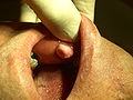 oral cavity fibroma
 -
 intramuscular lipoma
 -
 atrial myxoma
 -
 coccygeal chordoma
 -
 frontal sinus osteoma – CT
 -
 osteoid osteoma
 -
 Capillary hemangioma of the skin
 -
cavernous hemangioma in the liver
-
hemangioma
-
uterine leiomyoma
-
uterine leiomyoma
-
cardiac rhabdomyoma


.jpg)
Malignant mesenchymal tumors
Malignant mesenchymal tumors are termed as sarcomas (fish meat like appearance). They grow by infiltrative means and metastastizate mostly by hematogennous way. For the stage of malignity is decisive (more than the nuclear and cellular polymorphy) the frequency of mitosis, chromosomeal aberrations and presence and extent of necrosis. The material of intermediar filaments serves as a basic marker – vimentinor desmin in muscle cells (and parts of myofilaments).
Here belong:
- fibrosarcoma
- chondrosarkoma
- hemangiosarcoma
- leiomyosarkoma
- rhabdomyosarkoma
- osteosarkoma
-
Leptomeningeal fibrosarcoma
-
chondrosarcoma of the thoracic wall
-
Splenic hemangiosarcoma in a dog
-
uterine leiomyosarcoma
-
sclerosing rhabdomyosarcoma
Links
Realated articles
- Myxoma
- Lipoma
- Liposarcoma
- Hibernoma
- Hemangioma
- Angiosarcoma
- Lymfangioma
- Leiomyoma
- Leiomyosarkoma
- Rhabdomyoma
- Rhabdomyosarcoma
- Chondroma
- Chondrosarcoma
- Osteoma
- Osteosarcoma
Source
- https://www.wikiskripta.eu/w/Mezenchymov%C3%A9_n%C3%A1dory
- PASTOR, Jan. Langenbeck's medical web page [online]. [cit. 18.04.2010]. <https://langenbeck.webs.com/>.
- STŘÍTESKÝ, Jan. Patologie. 1. vydání. 2001. ISBN 80-86297-06-3.


.jpg)


.jpg)


.jpg)

.jpg)
