Liver
The liver is the largest exocrine gland in the body and a vital organ. Thanks to the large blood supply, they can serve primarily as a center for processing nutrients from food, a metabolic and detoxification center, a storage warehouse for glycogen, proteins and lipids. With their exocrine function, they ensure the excretion of bile, which helps in the digestion of fats. In the embryonic stage, they are also the seat of hematopoiesis.
Anatomy of the Liver[edit | edit source]
The liver has the shape of a three-dimensional triangle with a long hypotenuse from the lower right side to the upper left side. In an adult, they weigh on average around 1.5 kg and flow through them 1.5 l of blood/min. They are placed under the right part of the diaphragmatic arch and their end extends over the left medial part of the diaphragm. They press against the abdominal organs with their visceral surface (distinct impressions), their upper part is fused with the diaphragm.
- facies diaphragmatica - part of the liver attached to the diaphragm;
- facies visceralis - the lower part of the liver facing the abdominal organs.
These surfaces are separated in front by a sharp margo inferior, behind the visceral part passes into the diaphragmatic part without a sharp border.
Peritoneum forms a shiny coating on almost the entire surface of the liver - tunica serosa. Only on the diaphragmatic surface, where the f. diaphragmatica passes into the f. visceralis, is the area nuda, which is not covered by the serosa. tela subserosa attached to tunica fibrosa (capsula Glissoni), which is a firm and immovable covering of liver tissue. The tunica is related to the ligaments and blood vessels inside the liver.
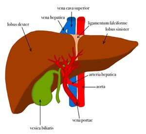
The liver is divided, like the lungs, into lobes:
- Right lobe - the largest liver lobe located on the right;
- Left lobe - smaller and flat left lobe;
- Quadrate lobe - quadrangular lobe in front between the right and left lobes, visible especially on the facies visceralis;
- Caudate lobe - caudate lobe at the back between the right and left lobes.
Segments of the liver:
The liver is divided into 8 functional segments. Thereby, the 3 vertically running hepatic veins, which together with their surrounding connective tissue are reffered to as fissures, divide the liver into four adjacent divisions.
The Divisio lateralis sinistra corresponds to the left anatomical lobe of the liver and reaches as far as the Lig. falcifome hepatis, behind which the left hepatic vein runs into the Fissura umbilicalis.
The Divisio medialis sinistra extends between the Lig. falciforme and the gall bladder, at which height the middle hepatic vein lies in the Fissura portalis principalis. Then to the right side follow the Divisio medialis dextra and the Divisio lateralis dextra, which are separated by the right hepatic vein in the Fissura portalis dextra but there is no visible landmark for this on the outer surface.These 4 vertical divisions are divided by the branches of the liver triad (V. portae hepatis, A. hepatica propria, Ductus hepaticus com- munis) into 8 liver segments.
- Segment I corresponds to the Lobus caudatus
- Segments II and III the anatomical left hepatic lobe
- Segment IV the Divisio medialis sinistra and segments V–VIII the rest of the anatomical right hepatic lobe, where the latter are numbered in a clockwise direction.
- The Lobus quadratus is a part of segment IVb.
- A ninth segment, which lies between segment VIII and I could be described, but is so far hardly taken into account in surgery.
In functional terms it is important that segments I–IV are supplied by the left branches of the liver triad and, therefore, in contrast to the macroscopically visible hepatic lobes, collectively belong to the left part of the liver (Pars hepatis sinistra), while segments V–VIII are dependent on the right branches of the liver triad and represent the functional right side of the liver (Pars hepatis dextra). Only segment I is regularly supplied from the branches of both sides.[1]
Impressions and Syntopy[edit | edit source]
1. Visceral surface:
o Gastric Impressions: the anterior wall of the stomach.
o Renal and Suprarenal impressions
o Duodenal impression: the ampulla of the duodenum
o Colic impression: the right colic flexure
o Esophageal impression: the abdominal part of the esophagus
2. Diaphragmatic surface:
o Cardiac impression: the heart in the middle of the diaphragmatic surface. The heart exerts pressure on left hepatic lobe (separated by the pericardium and diaphragm)
3. Anterior:
o Rib cage
o Anterior abdominal wall
4. Superior: Diaphragm
Facies visceralis[edit | edit source]
Especially on this side, the sagittal hepatic grooves - left and right and between them the transverse groove are visible. They separate the lobes and can be thought of as the letter H. The transverse depression is called the porta hepatis, which contains:
- entering a. hepatica propria (front left) and v. portae (back);
- emerging ductus hepaticus dexter et sinister (right and left bile ducts), it joins in the ductus hepaticus communis (front right).
Other important formations on this side include the fossa vesicae biliaris (storage of the gallbladder) at the right side of the lobus quadratus, where bile is stored and processed. The last prominent structure is the sulcus venae cavae laterally from the lobus caudatus, where thevena cava inferior runs. The inferior vena cava has either a transverse strip of lig. venae cavae, or is completely surrounded by liver tissue.
Position and projection of the liver[edit | edit source]
The liver is located in the diaphragmatic vault, touching:
- on the right lobe with adrenal gland, kidney, duodenum and with flexura coli dextra;
- on the left lobe with esophagus and stomach.
The organs leave their respective imprints on the f. visceralis.
the latter continues to the pars abdominis oesophagi and curvature minor of the stomach. It ends at the beginning of the duodenum in lig. hepatoduodenal.
The liver is projected in the regio hypochondriaca dextra (cartilages of the lower ribs on the right). Margo inferior begins at the edge of the right costal arch and continues to the medioclavicular line (the edge behind the 8th rib). From there, it proceeds obliquely to the left upwards (midway between the xiphoid process and the umbilicus) and ends behind the edge of the left costal arch, approximately midway between the edge of the sternum and the left medioclavicular line.
Liver fixation[edit | edit source]
Several mechanisms are involved in liver fixation. They are:
- League. teres hepatis - the rest after the umbilical vein, comes from the lig. falciform; fixes the liver to the anterior abdominal wall.
- Vena cava inferior - suspension of the liver to the VCI by means of the ligamentum venae cavae is an important "fixator" of the liver.
- Connection with diaphragm in the scope of area nuda - a part of the liver is connected to the diaphragm, which is referred to in Latin as pars affixa hepatis.
- Intestinal position - the organs located under the liver are also involved in the fixation of the liver, the liver "rests" on these organs.
- Intra-abdominal pressure
- The atmospheric pressure that stores the liver in the diaphragmatic vault also has a certain meaning. It can be broken by opening the abdominal cavity.
Histology of the liver[edit | edit source]
Hepatocyte[edit | edit source]
Polyhedral cell with dimensions of approx. 20–30 μm. Commonly contains one or two nuclei, which may be polyploid. It is a versatile cell with high metabolic activity. On the side facing the capillary (to Disse's space) it contains microvilli for a larger surface area for nutrient absorption.
The cell contains eosinophilic cytoplasm, mainly due to numerous mitochondria and smooth endoplasmic reticulum. This has several functions – notably methylation, oxidation and conjugation to modify xenobiotics before they are eliminated from the body.
Also present is rough endoplasmic reticulum for proteosynthetic activity. It forms clusters in the so-called basophilic bodies. Both proteins needed by the cell and blood serum proteins (ie albumins, prothrombins, fibrinogens, lipoproteins) are synthesized here. These are not stored, but escape directly into the bloodstream.
In coarse, electrodense granules, the cell stores liver glycogen, which is produced or degraded for the needs of the organism.
Other very important components of the hepatocyte include a high number of mitochondria (around 2,000), lysosomes, peroxisomes or Golgi complexes. The secretion of bilei is also essential.
Structure of liver parenchyma[edit | edit source]
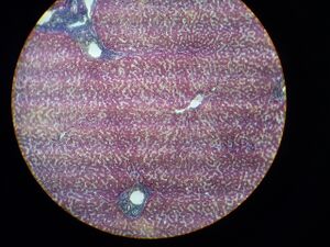
The morphological unit of the parenchyma is made up of liver cells, which create beams. Between these beams are the fenestrated sinusoids, where nutrients come from v. portae and oxygenated blood from a. hepatica propria. These beams, together with the vessels, form a radially arranged lobulus venae centralis with a central vein in the middle. It runs along the axis of the lobule and collects blood from the sinusoids, which it carries away from the liver.
The contact of the blood in the fenestrated capillary with the microvilli of the hepatocytes ensures the Disse's space (perisinusoidal) where the blood flows. On the lumina of the sinusoid there are also Kupffer cells (phagocytes) and infrequent Ito cells, which store lipid droplets, microfilaments, vitamin A in their cytoplasm and other important components. They also have a supporting function in liver regeneration.[2]
Bile ducts and triads[edit | edit source]
At the junction of two hepatocytes there is also a bile duct, the walls of which consist only of liver cell walls. It gradually passes into the ductus biliferi interlobulares. These leave in the portobiliary spaces, which contain three formations:
- arteria interlobularis from a. hepatica propria, which enters the lobule;
- vena interlobularis from v. portae, which also enters the lobule;
- ductus bilifer interlobularis, which emerges from the lobule.
This formation is collectively called trias hepatica (Glisson's trias)[3]. The vessels came here from the porta hepatis, when v. portae mostly accompanies a. hepatica propria. The ductus biliferi converge into a larger one and leave the port as the ductus hepaticus dexter et sinister.
Blood Flow[edit | edit source]
The functional flow of the liver is provided by the v. portae, which brings absorbed nutrients from the intestines. The nourishing component for the liver itself consists of the a. hepatica propria, which is one of the main branches of the truncus coeliacus. Nevertheless, it contributes little to the nutrition of hepatocytes with oxygen.
After entering through the porta hepatis, both vessels branch and form aa. et vv. interlobulares - part of the trias hepatica. In the portobiliary space, they also send out aa. et vv. circumlobulares' (distribution vessels) that surround the liver lobules.
In the trias hepatica, the vein joins the artery in the hepatic sinusoid between the hepatic trabeculae and continues to the center of the lobule in the v. centralis. They connect in the vena hepatis, which flows into the inferior vena cava in the sulcus venae cavae.
Primary hepatic acinus and portal lobule[edit | edit source]
The primary liver acinus is a functional liver unit, made up of two imaginary triangles that touch at their bases and have v. centralis at their apex. It is supplied by one circumlobular vein and artery. These send vessels to the sinusoids of two adjacent lobes. Primary hepatic acinus is histologically further divided into three zones:[4]
- zone I - the center of the liver acinus, it is closest to the circumlobular vein and artery, therefore there is the highest oxygen and nutrient supply;
- zone II - is further from the center of the liver acinus, smaller oxygen and nutrient supply;
- zone III - closest to the central vein, oxygen and nutrients arrive here as the last.
This division has a functional significance mainly for the examination of pathological conditions.
The portal lobule has peaks in three central veins centered in the portal triad. It thus occupies the functional parts of the three liver lobes.
Liver regeneration[edit | edit source]
Although the liver has a slow cell renewal, regenerative activity is high. Loss of liver tissue, whether from surgery or toxic agents, will prompt significant proliferation of cells. This results in almost complete replacement of the original tissue loss. It is probably caused by the so called chalons[5] that inhibit cell proliferation. If there are few cells, they also secrete few chalons and this leads to mitotic activity. In the case of many cells, enough chalons are secreted to suppress proliferation.
With repeated damage to the liver, there is a proliferation of ligament, which leads to irreversible changes.
Links[edit | edit source]
Related Articles[edit | edit source]
- Liver function
- Biochemical examinations of the liver • Diagnostic imaging methods in the examination of the pancreas, liver and spleen
- Hepatomegaly • Hepatosplenomegaly • Sarcoidosis of liver • Hepatic cysts and abscesses • Hepatic failure • Hepatic cirrhosis • Hepatitis • Liver Tumors • Liver Injury • Portal Hypertension
- Development of liver and gallbladder
- Liver (image) • Liver - HE • Liver (SFLT) • Liver - PAS • Chronic liver abscess (slide) • Focused large droplet steatosis of the liver (slide)
- Biliary tract • Gallbladder • Spleen • Kidneys
Sources[edit | edit source]
- PASTOR, Jan. Langenbeck's medical web page [online]. [cit. 2009]. <http://langenbeck.webs.com>.
Bibliography[edit | edit source]
- ŠIHÁK, Radomír – GRIM, Miloš. Anatomy 2. Second edition. Prague : Grada, 2002. 488 pp. pp. 127-138. ISBN 80-247-0143-X.
- JUNQUIERA, L. Carlos – CARNEIRO, José – KELLEY, Robert O, et al. Základy histologie. 1. edition. Jinočany : H & H, 1997. 502 pp. pp. 303-317. ISBN 80-85787-37-7.
References[edit | edit source]
- ↑ SOBOTTA, Sobotta Anatomy Textbook, Elsevier GmbH, Munich, Germany. 2019 ISBN 978-3-437-44080-9
- ↑ CHIHÁK, Radomír – GRIM, Miloš. Anatomy 2. Second edition. Prague : Grada, 2002. 488 pp. pp. 134. ISBN 80-247-0143-X.
- ↑ CHIHÁK, Radomír – GRIM, Miloš. Anatomy 2. Second edition. Prague : Grada, 2002. 488 pp. pp. 135. ISBN 80-247-0143-X.
- ↑ JUNQUIERA, L. Carlos – CARNEIRO, José – KELLEY, Robert O, et al. Základy histologie. 1. edition. Jinočany : H & H, 1997. 502 pp. pp. 315. ISBN 80-85787-37-7.
- ↑ JUNQUIERA, L. Carlos – CARNEIRO, José – KELLEY, Robert O, et al. Základy histologie. 1. edition. Jinočany : H & H, 1997. 502 pp. pp. 315. ISBN 80-85787-37-7.


