Kidneys
From WikiLectures
The kidneys are made up of a tubular gland , where each tubule is called a nephron . The nephron is also functionally connected to the collecting and draining ducts.
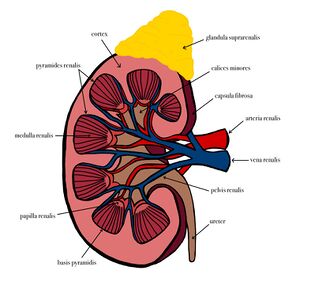
The kidney has a specific bean-like shape, where we can distinguish:
- 'Facies anterior' ,
- 'Facies posterior' ,
- 'Extremitas Superior' ,
- 'Extremitas inferior' ,
- 'Margo lateralis et medialis.
On medial margin we find a packed place, Renal hilum.
The dimensions of the kidney are (length) 10–12 cm X (width) 5–6 cm X (thickness) 3.5–4 cm. One kidney is able to perform its function in case of defect of the other kidney.
Structure of the Kidney[edit | edit source]
See the Kidney function page for more detailed information
Function[edit | edit source]
- Primary urine formation and adjustment to definitive urine (hypertonic);
- Urine excretion (mainly urea), metabolites;
- maintenance of homeostasis, regulation of acid-base balance;
- Endocrine function (renin, erythropoetin, 1,2-dihydroxycholecoferol);
- Regulation of water volume in the body;
- Excretion of toxic substances, medicines and other metabolites;
- Regulation of blood pressure.
Regulatory mechanisms[edit | edit source]
- Autonomous nerves (mainly effect on blood vessel contractility);
- Endocrine regulation (the basis is juxtaglomerular apparatus).
Compensatory mechanisms[edit | edit source]
- Myogenic autoregulation - the blood vessel responds to a change in pressure - when the pressure drops, the vas afferens dilates and when the pressure increases, vasoconstriction occurs. Myogenic regulation is mediated by endothelins.
- Renin - aldosterone - angiotensin system - this includes the cells of the juxtaglomerular apparatus ( JGA ) of the kidneys. It is made up of three types of cells:
- juxtaglomerular cells;
- macula densa cells – register the change in ion concentration;
- juxtaglomerular mesangial cells . JGA produces renin (the stimulus for its synthesis is a decrease in pressure in the vas afferens or a decrease in ion concentration ), which enables the conversion of angiotensinogen to angiotensin I , which is transformed into angiotensin II due to ACE (angiotensin-converting enzyme) . It has vasoconstrictive properties mainly in the vas efferens. In addition, it also stimulates the production of aldosterone (ALD) .
- it affects the filtration fraction – the filtration fraction is the ratio between glomerular filtration and plasma flow in the kidneys (physiologically approx. 15-20% ).
The blood vessels of the kidneys[edit | edit source]
- Renal circulation accounts for about 20% of cardiac output —this large flow is of greater functional importance than the circulation that supplies nutrients to the kidneys. The high pressure in the vas afferens exceeds the oncotic pressure throughout the glomerulus.
- Skimming effect – separation of erythrocytes from plasma. Erythrocytes pass through wider sinuses and plasma is filtered by capillaries.
- Arteries
- A. renalis dextra et sinistra – each is divided into anterior branches (4 branches for the anterior segments) and posterior branch (for the back segment).
- A. renalis accesoria - occurs in 30%.
- Branching for the cortex: arteriae lobares, arteriae interlobares, arteriae arcuatae, arteriae interlobulares - of them arteriae glomerulares afferentes/efferentes, peritubular capillary bed.
- Branching for the medulla: the basis is arteriae glomerulares efferentes, then arteriae rectae.
- Venous outflow
- Veins from the cortex: venulae stellatae – peritubular capillary bed – venae interlobulares – venae arcuatae – venae interlobares – vena renalis.
- Veins of the pulp: venulae rectae – venae arcuatae.
- Lymph
- Lymph collects in three braids, from the peritubular, subcapsular, and capsular adiposa spaces , and arrives at the lumbar lymph nodes .
- Nerves
- it comes from the renal plexus = a plexus of sympathetic, parasympathetic and sensitive fibers passing around the a. renalis . Autonomic fibers have a vasomotor function, sensitive fibers mainly innervate the capsula fibrosa , so that the parenchyma itself is practically insensitive.
- Arteries
Position and fixation of kidneys[edit | edit source]
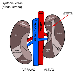
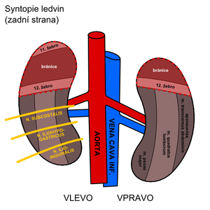
- They are located in the retroperitoneal space at the height of Th12–L2, the upper third lies on the diaphragm, the lower two thirds on the quadratus lumborum muscle ;
- The right one is shifted about half the length of the vertebra lower;
- The renal hilum projects skeletotopically to the level of L1.
Links[edit | edit source]
Related articles[edit | edit source]
- Kidney (histological preparation)]
- Nephron
- Kidney function in maintaining acid -base balance
- Tubular processes
- Blood flow through the kidneys and its self -regulation
External links[edit | edit source]
- MAREKOVÁ, Maria – ORAVSKY, R., et al. Multidisciplinary Putting to Facials [online]. Portal of the Faculty of Lecture of Pavel Jozef Šafárik University in Košiciach, ©2011. The last revision 2011-11-25, [cit. 2011-11-25]. <https://portal.lf.upjs.sk/clanky.php?aid=134>.
Used literature[edit | edit source]
- ČIHÁK, Radomir – GRIM, Milos. anatomy. 2nd, mod. and the add -on edition. Prague : Grada Publishing, 2002. 470 pp. ISBN 80-7169-970-5.
- GANONG, William f. overview of medical physiology. 20. edition. Prague : Galen, 2005. 890 pp. ISBN 80-7262-311-7.
- TROJAN, Stanislav, et al. medical physiology. 4. edition. Prague : Grada, 2003. 772 pp. ISBN 80-247-0512-5.


