Kidney
The kidneys are responsible for the removal of excess water, salts and wastes of protein metabolism from blood and at the same time retaining nutrients and chemicals to the blood.
Structure
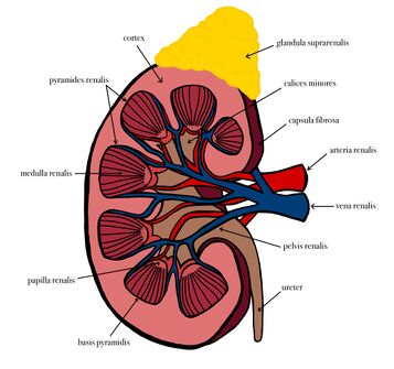
They are bean-shaped, convex lateral margin, concave medial margin. Their color is reddish-brown and has measurements about: 10cm length, 5cm width, and 3cm thickness. The renal hilum (portal renalis) lies in the medial margin, a space where blood vessels, nerves and the renal pelvis (and its adipose tissue) enter and leave.
The kidneys are contained into the renal capsule; a tough capsule made of collagenous fibers and connected on the kidney by areolar tissue. As it proceeds medially towards the hilum, it connects with the connective tissue of the vessels which are entering a central recess, the renal sinus.
They have superior and inferior poles. On the superior poles, there are located the suprarenal glands. They are also enclosed in a capsule and in a cushion of pararenal fat. They are separated from the kidneys by a weak fascial septum, thus the 2 structures are not actually attached to each other.
Syntopy
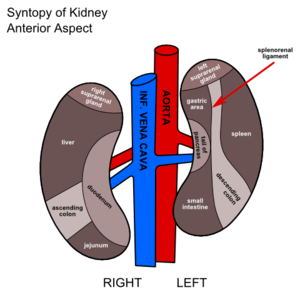
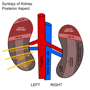
The right kidney is lower than the left by 1 rib difference. This is because of the prominence of the inferior side of the liver on the right side of the body. The right kidney is separated from the liver by the hepatorenal recess. Perirenal fascia (or renal fascia envelope), encloses the kidneys and the suprarenal glands and contain the perirenal fat as cushioning for these structures. The anterior part is called prerenal fascia and the posterior retro-renal fascia. Pararenal fat is located behind the retrorenal fascia and the fascia of quadratus lumborum.
== Cortex ==
It is brownish-red in color, 1 cm wide and lies beneath fibrous renal capsule. Medullary rays penetrate the cortex, starting from the base of the pyramids (medulla), forming the pars radiate of the cortical lobules. In those lobules, the partes convulutae are located. The dark red spots are the renal corpuscle (glomeruli), the site of ultrafiltration. Between the pyramids of the medulla, the cortical substance lies in the form of renal columns, that extend down to the renal pelvis.
Medulla
It consists of several large pyramids. Their apices (papillae) point towards the renal pelvis. It can be distinguished in 2 zones:
- the reddish external zone (with internal and external striations);
- the pale internal zone.
The medulla contains the straight vessels, arteriolae and venulae rectae which reabsorb the nutrients from the primary urine (formed in the cortex by ultrafiltration) back to the bloodstream.
Envelopes
The kidneys are contained into the renal capsule; a tough capsule made of collagenous fibers and connected on the kidney by areolar tissue. As it proceeds medially towards the hilum, it connects with the connective tissue of the vessels which are entering a central recess, the renal sinus. Perirenal fascia (or renal fascia envelope), encloses the kidneys and the suprarenal glands and contain the perirenal fat as cushioning for these structures. The anterior part is called prerenal fascia and the posterior retro-renal fascia. Pararenal fat is located behind the retrorenal fascia and the fascia of quadratus lumborum muscle.
Renal vessels arise at the level of the intervertebral disc between L1 and L2 vertebrae.
Renal arteries and their branches

The arteries are mostly posterior to the veins. They divide close to the hilum into 5 segmental arteries, which are end-arteries (they do not anastomose significantly with adjacent branches). The right renal artery is the longest vessel and passes also posterior to inferior vena cava. Before entering the hilum, the renal artery gives 2 branches:
- the inferior suprarenal artery;
- ureteric branch.
The segmental arteries of the renal arteries:
- Superior (apical) (from anterior branch of renal aa)
- Anterior superior (from anterior branch of renal aa)
- Anterior inferior (from anterior branch of renal aa)
- Inferior (from anterior branch of renal aa)
- Posterior (continuation of the posterior branch of renal aa)
The segmental arteries then subdivide in the following manner:
- Interlobar arteries in the renal sinus between the pyramids →
- Arcuate arteries that go around the pyramids in the medulla region →
- Cortical radiate arteries that “radiate” around the pyramids into the cortical region. Some of these can also perforate the renal capsule.
- Arcuate arteries that go around the pyramids in the medulla region →
Renal veins and their tributaries
Veins are numerous and drain in each kidney in a variable fashion to form the renal vein. The renal veins both lie anterior to the renal arteries. The left renal vein, receives also:
- the testicular (ovarian) vein;
- the left suprarenal vein;
- communicating branch with ascending lumbar vein.
Lymphatics
Lymph is conducted along lymphatic vessels that follow the renal veins and drain into the right and left lumbar (caval & aortic) lymph nodes.
==Renal sinus description & syntopy==
The renal sinus, which lies inside the hilum of the kidney, contains the renal pelvis and its calices, together with the other structures, nerves, vessels and fat.
Pelvis
The renal pelvis is the flattened, funnel-shaped expansion of the superior end of the ureter and its apex is continuous with the ureter. The pelvis receives 2–3 major calices, each of which sub-divides into 2–3 minor calices. The pelvis can exist as wide ampulla-shaped or tubular (branched). In living persons, the pelvis and its subsequent calices are collapsed (empty).
Calyx
Each minor calyx surrounds a renal papilla (apex of pyramid) and collects the urine.
Ureter
Ureter is shaped like a slightly flattened tube of about 4–7 mm diameter. It has variable length, but average might be around 30 cm. It is about 1 cm shorter in the female.
Syntopy of ureter
- Runs inferiorly from the apex of the renal pelvis at the hilum.
- Passes over the pelvic grim at the bifurcation of the common iliac artery.
- Runs along the lateral wall of pelvis and enters the urinary bladder.
- Abdominal parts adhere closely to the parietal peritoneum and are retroperitoneal throughout their course.
- Three constrictions:
- At junction between renal pelvis and ureter.
- At crossing of brim of pelvic inlet.
- During passage through wall of urinary bladder.
Vasculature of ureter
- Abdominal portion: branches from renal arterties, abdominal aorta and common iliac artery.
- Pelvic portion: branches from superior vesical aa, middle rectal aa, uterine/vaginal (female), inferior vesical artery (male).
Veins: follow the arteries in the same way and drain into renal and testicular (ovarian) veins.
Kidney structure:
Kidney functions:
1. Purification (pH, ions like K+, urea, creatinine):
2. Synthetic function (↑glucose, EPO, TPO, calcitriol):
3. Degradation/Deactivation (PTH, insulin):
Links
Related articles
- Renal Blood Vessels and Renal Segments
- Renal Pelvis and Calices
- Structure of Kidney
- Ureter
- Urinary bladder
Bibliography
- MOORE, Keith L – DALLEY, Arthur F. Clinically Oriented Anatomy. 5. edition. Lippincott Williams & Wilkins, 2005. ISBN 0781736390.


