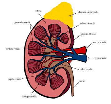
Structure of Kidney
Cortex
It is brownish-red in color, 1 cm wide and lies beneath fibrous renal capsule. Medullary rays penetrate the cortex, starting from the base of the pyramids (medulla), forming the pars radiate of the cortical lobules. In those lobules, the partes convulutae are located. The dark red spots are the renal corpuscle (glomeruli), the site of ultrafiltration. Between the pyramids of the medulla, the cortical substance lies in the form of renal columns, that extend down to the renal pelvis.
Medulla[edit | edit source]
It consists of several large pyramids. Their apices (papillae) point towards the renal pelvis. It can be distinguished in 2 zones:
- the reddish external zone (with internal and external striations);
- the pale internal zone.
The medulla contains the straight vessels, arteriolae and venulae rectae which reabsorb the nutrients from the primary urine (formed in the cortex by ultrafiltration) back to the bloodstream.
Envelopes[edit | edit source]
The kidneys are contained into the renal capsule; a tough capsule made of collagenous fibers and connected on the kidney by areolar tissue. As it proceeds medially towards the hilum, it connects with the connective tissue of the vessels which are entering a central recess, the renal sinus. Perirenal fascia (or renal fascia envelope), encloses the kidneys and the suprarenal glands and contain the perirenal fat as cushioning for these structures. The anterior part is called prerenal fascia and the posterior retro-renal fascia. Pararenal fat is located behind the retrorenal fascia and the fascia of quadratus lumborum muscle.
Links[edit | edit source]
Related articles[edit | edit source]
Bibliography[edit | edit source]
- MOORE, Keith L – DALLEY, Arthur F. Clinically Oriented Anatomy. 5. edition. Lippincott Williams & Wilkins, 2005. ISBN 0781736390.

