Bone
Bone (os) is mineralized connective tissue that is formed by a process called ossification. It is created by the activity of osteoblasts, which are cells that produce bone matrix. After their incorporation into the bone tissue, they are called osteocytes. All the bones together form the skeleton, which is the basis of the passive locomotion apparatus. Muscles and ligaments attach to the skeleton. The entire surface of the bone (except for the places where muscles/ligaments attach to the bone and the joint heads, which are covered by cartilage) is covered by a fibrous covering of the periosteum, the inner surfaces by the endosteum. There are about 207 bones in our body. (Number changes with age)
Bone composition[edit | edit source]
Bone matrix has two basic components:
- Constituent – 1/3 organic component, also called ossein. It is made up of collagen fibrils and an amorphous mass, which has osteoalbumoid and osteomucoid as its basic components.
- Non-structural – 2/3 salt crystals (calcium phosphate and hydroxyapatite) that are built into the structural component.
Individual components can be separated from each other - either by leaching in weak acids (the mineral salts are removed and the bone becomes very soft) or by annealing (this removes the structural component and the bone becomes very fragile).
Bone structure[edit | edit source]
2 main types of bone tissue are involved in bone building:
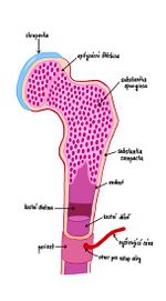
- Dense bone tissue (substantia compacta) – it is located on the surface of the bone. Its structure is arranged in layers - lamellae (lamella bone). These lamellae are typically arranged in cylindrical formations - osteons (Haversian systems). An osteon can form up to 20 lamellae concentrically arranged around a central canal. Osteocytes are located between the lamellae.
- Spongy bone tissue , bone beam (substantia spongiosa) - located inside the bone.
Bone tissue remodeling takes place throughout life, slower in old age. Bones also change their structure according to the direction of the load (e.g. beams from spongy bone tissue in the head of the femur).
Dividing bones[edit | edit source]
According to the form[edit | edit source]
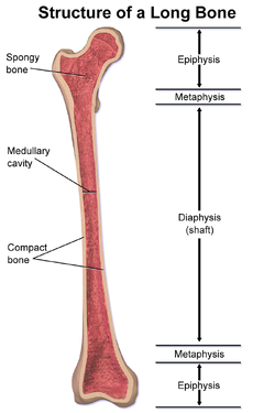
- Long bones - we distinguish epiphyses (epiphysis) on them, which are either on one or both ends of a long bone. Among them is the diaphysis (diaphysis), which corresponds in its extent to the body of the bone. Furthermore, during the growth period, there are growth (epiphyseal) cartilages at the transition between the body and the heads, thanks to which the bone increases in length. The surface of the bone is formed by a thick layer of compact tissue on the diaphysis, whereas the epiphyses are covered with only a weak layer of compact tissue. The inside of the bone is made up of cancellous tissue. The cavity in the body of the bone (cavitas medullaris) is filled with bone marrow (medulla ossium). The ossifying nuclei of a long bone are:
- In the diaphysis – in the center of the bone, from where ossification progresses to the ends;
- In the pineal glands;
- Alternatively, additional ossification nuclei (apophyses) may occur in large bone bumps, or in places where muscles are attached - e.g. in the greater trochanter of the femur (trochanter major).
- Short bones - on their surface there is a thin layer of compacts (substantia corticalis), inside they are filled with spongiosa. Ossification of a short bone always takes place enchondrally and from one ossification core, which is approximately in the center of the bone, from where it progresses to the surface throughout the growth period.
- Flat bones - these bones always have a layer of compacta (lamina externa, lamina interna) on the outer and inner surfaces, between which there is a layer of spongiosa. Flat bones ossify from multiple nuclei.
According to the method of ossification[edit | edit source]
- Desmogenous ossification – bones arising from ligaments (e.g. some bones of the skull, clavicle).
- Chondrogenic ossification – the original cartilaginous model of the bone is replaced by bone tissue (e.g. humerus).
Bones pneumatized[edit | edit source]
Pneumatized bones (ossa pneumatica) are bones into whose spongiosa the mucous membrane of the nose or middle ear cavity began to protrude after birth. These cavities remain connected to the original cavity by a narrow passage.
Bone vessels and nerves[edit | edit source]
Bone vessels separate from surrounding large arteries or merge into surrounding veins. Alternatively, they are separated/poured into the vascular networks, which are usually on the joints.
Arteries[edit | edit source]
- Long bones
- The diaphysis of a long bone is supplied in several ways:
- Arteria nutricia – one or two thicker arteries.
- Vessels from the periosteum - a large number of vessels that enter the bone through Volkmann's canals and connect to the vessels in the Haversian canals.
- Arteriae metaphysariae – vessels supplying the expanding end of the bone at the junction between the diaphysis and the epiphysis. They mostly come out of joint knitting.
- The epiphyses are supplied through the arteriae epiphysariae , which separate from the articular networks.
- The diaphysis of a long bone is supplied in several ways:
- Short bones - their supply is from surrounding vessels or networks. The vessel enters the bone on the surface facing the joint capsule.
- Flat bones - they are supplied by wide arteriae nutriciae and a greater number of periosteal arteries.
Bone veins[edit | edit source]
Veins drain blood along arteries, possibly through separate channels, and enter surrounding veins or networks.
Nerves[edit | edit source]
Nerves enter the bone from the innervated periosteum through the Haversian canals.
Links[edit | edit source]
Virtual microscope[edit | edit source]
Histologie.png Enchondral Ossification - HE (big toe)
Histologie.png Desmogenous ossification - HE
Histologie.png Lamellar bone compact - HE (finish)
Histologie.png Cancellous lamellar bone - HE
Related articles[edit | edit source]
- Ossification
- Chondrogenic ossification
- Desmogenous ossification
- Bone growth and healing
- Bone structure and remodeling
- Microscopic structure of bone tissue
- Cartilage
- Ligament
- epithelium
External links[edit | edit source]
References[edit | edit source]
- ČIHÁK, Radomír – GRIM, Miloš. Anatomie. 2. upr. a dopl edition. Praha : Grada Publishing, 2001. vol. 1. ISBN 80-7169-970-5.


