Connection of bones
A bone junction (juncture) is where two or more bones meet. It is used to move a certain part of the body. The science of bone connections is called arthrology or syndesmology.
Bone Connection Types[edit | edit source]
There are two types of bone connections:
- Smooth joint - bones are connected by a binder (ligament, by cartilage or bone).
- Connection by touch - bones touch each other in places known as contact surfaces, on the sides of these surfaces there is a ligament that also connects the bones. This connection is referred to as joint joint (junctura synovialis) - joint (articulatio).
Connection smooth[edit | edit source]
Juncture fibrosa[edit | edit source]
Junctura fibrosa is the connection of bones by ligaments. It is used where tensile stress occurs. We can divide it into other types:
- Syndesmosis are bones connected by ligaments outside the skull. The strip of tissue that connects the bones in this way is a ligament (ligamentum).
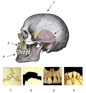
- Suture (seam) is the connection of flat bones skulls by ligament. We distinguish several types according to shape:
- Sutura serrata (saw-shaped seam): the edges of adjacent bones press against each other with saw-shaped protrusions (in the entire thickness of the bone), which expands the contact surface of the bones and thus increases the strength of the connection.
- Squamosa suture (scaly suture): the thinned edge of one bone is placed over the edge of the other bone, thereby widening the surface of the fibrous connection.
- Sutura plana (smooth seam): straight, smooth edges of adjacent bones are joined by ligaments. It is most often found in places that are not mechanically stressed.
- Gomphosis (junctura dentoalveolaris) is the junction of a tooth and a socket in the jaw. The tooth is attached by a ligament called the periodontium.
Junctura cartilaginea[edit | edit source]
It is the connection of bones with cartilage. This is a fixed connection. Again, we can divide it into other types:
- Synchondrosis is a combination where hyaline cartilage predominates. An example is the connection of the bones sternum.
- 'Symphysis is a firm and flexible connection, which was created due to alternating stress in compression and tension. Fibrous cartilage predominates here, only a small proportion close to the bones is hyaline cartilage. An example is pubic symphysis.
Osseous juncture[edit | edit source]
Also synostosis' - joining of two bones with the help of a bone. It develops secondarily from the fibrous or cartilaginous connection of bones by desmogenous and chondrogenic ossification. An example is the union of the sacral vertebrae in the sacrum.
Touch Connection[edit | edit source]
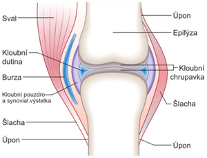
A joint (articulatio) is a movable connection of two or more bones that touch surfaces covered with cartilage inside a fibrous capsule. The contact surfaces (facies articulares) usually have the shape of the articular fossa (fossa articularis), which is concave, and the articular head (caput articularis), which is convex. The fibrous articular capsule (capsula articularis) connects the bones around the perimeter of the contact surfaces, while in individual joints it is attached at different distances from the edges of the cartilaginous contact surfaces. It is loose enough to allow extreme deflections of the bones during movements. The joint cavity (cavitas articularis) is a gap between the contact surfaces, the capsule and other structures in the joint, filled with synovial fluid (which is produced by the joint capsule). The joint may include special devices:
- Articular rim (labrum articulare) is the raised edge of the articular surface formed by fibrocartilage, it widens the area of the articular fossa.
- Articular discs and menisci are plates of fibrocartilage that are inserted between the joint surfaces. Discus articularis has the shape of a solid target and divides the joint into two cavities. The meniscus articularis has a sickle shape, it is higher on the outside and flattens towards the contact surfaces. Their purpose is to compensate for the uneven curvature of the surfaces of the socket and the head, and they also enable more complex movements in the joint.
- Articular ligaments strengthen the joint capsule and influence its movements. They can be built directly into the capsule (ligamenta capsularia), or attached to the surface of the joint and separated by a ligament (ligamenta extracapsularia), or they are directly inside the joint (ligamenta intracapsularia).
- Bulky bursae (bursae synoviales) are found in the thin connective tissue around the joint. They are cavities lined with a synovial membrane and contain a fluid similar to synovial fluid. They occur in places where tendons or ligaments rub against the joint capsule.
- Musculi articulares are small muscles that are attached to the joint capsule from the deepest layers of the surrounding muscles and during movement by pulling on the capsule, they prevent it from getting stuck between the joint surfaces.
Split joints[edit | edit source]
According to the number of components[edit | edit source]
- Simple joint (articulatio simplex) in which it articulates she only eats two bones.
- Compound joint (articulatio composita) in which more than two bones or two bones and some special joint device meet.
According to the shape of the contact surfaces[edit | edit source]
- spheroidal (a. spheroidea) - Both the head and the socket are parts of the sphere's surface. Movement is possible in combination along three mutually perpendicular axes.
- free (arthrodia) - The area of the socket is smaller than the area of the head, allowing a large range of motion. E.g. articulatio humeri.
- restricted (enarthrosis) – The socket is deep, so the head rests against it, causing less range of motion. E.g. hip joint
- ellipsoidal (a. ellipsoidea) – The contact surfaces are similar to a rotating ellipsoid. Movement is possible in two directions: around the long axis, sliding bows of the head to the sides. E.g. articulatio atlantooccipitalis, articulatio bicondylaris (mandible at the base of the skull)
- saddle (a. sellaris) - The contact surfaces are in the shape of a horse saddle with a rider sitting in it. Movement is possible in two directions and in their combination.
- cylindrical (a. cylindrica) – The contact surfaces are in the shape of a cylinder.
- articular (ginglymus) – The axis of movement is perpendicular to the longitudinal axis of the bone. E.g. articulationes interphalangeales
- wheel (a. trochoidea) – The joint has the shape of a low cylinder and its axis of rotation is identical or parallel to the longitudinal axis of the bone. E.g. articulatio radioulnaris proximalis et distalis
- pulley (a. trochlearis)' - The basic shape of the joint is cylindrical, but on one of the joint surfaces (mostly on the head) it has a guide groove, so the head has the shape of a pulley, then into it the guide bar emerging from the second joint surface fits into place. There are no sideways movements. E.g. articulationes interphalangeales, articulatio humeroulnaris
- flat (a. plana) – In this joint, the contact surfaces are almost flat and slide over each other during movement. It usually has strong joint ligaments that limit movement. E.g. articulatio acromioclavicularis
- stiff (amphiarthrosis) – A joint similar to a flat joint, but with irregular and slightly wavy contact surfaces, so movement is even more limited (joint with slight sliding mobility). E.g. articulatio iliosacralis
Links[edit | edit source]
Related Articles[edit | edit source]
References[edit | edit source]
- ČIHÁK, Radomír. Anatomie I. 2. edition. Praha : Grada, 2001. 516 pp. ISBN 978-80-7169-970-5.


