Blood vessels
Blood vessels are part of the lymphatic and cardiovascular systems. Through the cardiovascular system, oxygen and nutrients are distributed in the body to the tissues and waste products of metabolism to the excretory organs. Furthermore, the transport of hormones to the target organs is mediated. Using the lymphatic vascular system, fluid from the intercellular spaces returns to the blood circulation. These systems thus contribute to the integration of the function of the entire organism.
Vessels are divided into blood vessels and lympatic vessels.
In blood vessels, we further distinguish arteries, veins and capillaries.
General structure of blood vessels[edit | edit source]
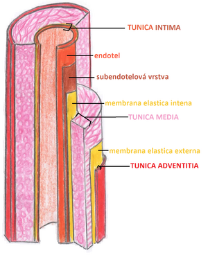
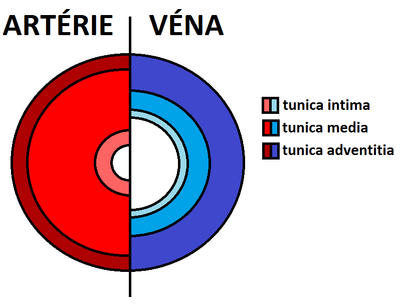
Blood vessels are composed of three basic structures: tunica intima, tunica media and tunica adventitia.
Tunica intima[edit | edit source]
It consists of a layer of endothelial cells abutting the basal lamina and a subendothelial layer.
Endothelial cells[edit | edit source]
- Polygonal, flat;
- stretched in the direction of blood flow;
- the central region arches into the lumen of the vessel;
- they have thin lateral projections - they often contain pinocytic vesicles (for the transport of substances).
Lamina basalis[edit | edit source]
- Endothelial cell product;
- may or may not be continuous.
Subendothelial layer[edit | edit source]
- Thin callagenous tissue;
- may contain smooth muscle cells;
- may contain smooth muscle cells;
Tunica media[edit | edit source]
This layer is formed by smooth muscle cells, that produce intercellular mass (glycosaminoglycans, chondroitin sulfate and proteoglycans). We also find reticular fibers and elastic fibers here. At the edges, they can condense in the membrana elastica interna et externa (they separate the tunica media from the tunica externa and tunica intima).
Tunica adventitia[edit | edit source]
The layer is made up of collagen fibers, in which longitudinal collagen (primarily collagen I ) and elastic fibers predominate. We also find fibroblasts, adipocytes and smooth muscle cells in larger vessels.
Nutrition of blood vessels[edit | edit source]
Nutrition of blood vessels[edit | edit source]
The nutrition of the wall of small vessels is provided by the diffusion of nutrients and oxygen from the blood flowing inside the given vessel. Vessels that are larger than 1 mm in diameter have their own vascular system developed in the walls. This system is called the vasa vasorum. Vasa vasorum arise as branches of one's own artery or a neighboring artery. These vessels branch in the tunica adventitia and in the outer regions of tunica media. Because there is less oxygen concentration in venous bloode vasa vasorum occur more often in the walls of veins than in the walls of arteries.
Lymphatic drainage[edit | edit source]
Lymphatic capillaries are found mainly in the tunica adventitia of vessels. In the veins, they penetrate deeper (up to the tunica media).
Vasomotor innervation[edit | edit source]
A network of vasomotor nerve fibers (sympathetic unmyelinated) is found in the walls of most blood vessels, which contain smooth muscle cells. Their chemical mediator is noradrenaline, which when released causes vasoconstriction. In arteries, nerve fibers usually do not penetrate the tunica media, noradrenaline has to diffuse several micrometers to reach the smooth muscle cells of the tunica media. In veins, we find nerve endings in the tunica adventitia and in the tunica media, but the total number of nerve endings is smaller than in arteries.
Links[edit | edit source]
Relatide Articles[edit | edit source]
References[edit | edit source]
MESCHER, Anthony L. Junqueira's Basic Histology. 12. edition. United States : McGraw-Hill Education - Europe, 2009. 480 pp. ISBN 9780071630207.
KONRÁDOVÁ, Václava – UHLÍK, Jiří – VAJNER, Luděk. Funkční histologie. 2. edition. Jinočany : H & H, 2000. 291 pp. ISBN 80-86022-80-3.
PAULSEN, Douglas F. Histologie a buněčná biologie : Opakování a příprava ke zkouškám. 1. edition. Jinočany : H & H, 2004. 433 pp. ISBN 80-7319-024-9.


