Bone marrow
__CONTENT__
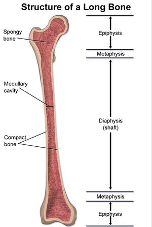
The bone marrow (medulla ossium) is a hemopoietic organ from the 7th month of the prenatal period (the so-called medullo-lymphatic period). It is found in the bodies of long bones and in the spaces between the trabeculae of spongiosa. It fills the medullary cavity (cavitas medullaris). A soft, macroscopically diverse tissue composed of reticular tissue. A sternal puncture is obtained for microscopic examination.

Medulla ossium rubra = red bone marrow[edit | edit source]
It is a hematopoietic organ. It consists of a spatial network of reticular tissue (reticular cells, fibers), macrophages and wide blood capillaries. There are stem cells for the formation of erythrocytes, most leukocytes and platelets in the meshes of the ligament. Red bone marrow remains in the spongiosa of the articular ends of long bones, in the spongiosa of short bones, in the pelvic bones, in the sternum, ribs, and in the diploe of the cranial bones.
Contains:
- reticular cells - form a network in which stem cells are located;
- fat cells - their quantity can change depending on the need for blood formation;
- macrophages'
- phagocytose apoptotic immature blood cells and old erythrocytes
- participate in the control of hemopoiesis by releasing factors
Medulla ossium flava = yellow bone marrow[edit | edit source]
It arises from red bone marrow. In the marrow of long bones, hematopoiesis ceases during growth, the reticular ligament is penetrated by fat cells, and the red marrow turns yellow. By age 20, yellow bone marrow is in the cavities of all long bones (exception - the proximal end of the body of the humerus and femur). It is capable of reactivation to red bone marrow.[edit | edit source]
Medulla ossium grisea = gray bone marrow.[edit | edit source]
- It is translucent and arises from the yellow pulp through the loss of fat. It is typical for late age.
Bone vessels and nerves[edit | edit source]
Bone vessels separate from surrounding large arteries or merge into surrounding veins. Alternatively, they are separated/poured into the vascular networks, which are usually on the joints.
Arteries[edit | edit source]
. Long bones : The diaphysis of a long bone is supplied in several ways:
-Arteria nutricia – one or two thicker arteries.
-Vessels from the periosteum - a large number of vessels that enter the bone through Volkmann's canals and connect to the vessels in the Haversian canals.
-Arteriae metaphysariae – vessels supplying the expanding end of the bone at the junction between the diaphysis and the epiphysis. They mostly come out of joint knitting.
. The epiphyses are supplied through arteriae epiphysariae, which separate from the articular networks.
. Short bones : their supply is from surrounding vessels or networks. The vessel enters the bone on the surface facing the joint capsule
. Flat bones : they are supplied by wide arteriae nutriciae and a greater number of periosteal arteries.
Nerves[edit | edit source]
Nerves enter the bone from the innervated periosteum through the Haversian canals.
Links[edit | edit source]
Related Articles[edit | edit source]
References[edit | edit source]
- ČIHÁK, Radomír. Anatomie 1. 2. edition. Grada, 2001. 497 pp. ISBN 80-7169-970-5.
- LÜLLMANN-RAUCH, Renate. Histologie. 3. edition. Grada, 2012. 576 pp. ISBN 978-80-247-3729-4.


