The Third Week of Embryo Development
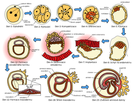
The third week of development is characterized by the emergence of the primitive streak , the development of the notochord and the differentiation of the three germ layers (gastrulation).
Gastrulation[edit | edit source]
Gastrulation is the process by which a two-layered target changes into a three-layered one. It begins with the formation of a primitive streak on the surface of the epiblast. Each of the terminal cotyledons gives rise to certain tissues:
| Germ leaf | Derived tissue |
|---|---|
| ectoderm | skin and its derivatives (eyelashes, hair, hair, nails), CNS and PNS, retina, etc. |
| endoderm | respiratory system (larynx, trachea, lungs), digestive system outside the oral cavity and anus (liver, gallbladder, pancreas, stomach, small and large intestine) |
| mesoderm | muscles, connective tissue, cardiovascular system (heart, vessels, blood), lymphatic system, bones, reproductive and excretory system |
Primitive Strip[edit | edit source]
At the beginning of the third week, a distinct opacity formed by a thickened band of epiblast appears caudally in the mid-plane of the dorsal surface . It arises from the proliferation and migration of epiblast cells towards the medial plane of the target. The strip elongates caudally and the anterior end proliferates to form a primitive node. At the same time, we observe that a longitudinal primitive furrow is formed in the axis of the strip , which connects to the depression in the node in the front - a primitive pit. Both the pit and furrow are the result of invagination of epiblast cells.
- Cells departing from the primitive streak form
- between the epiblast and the hypoblast, a thin network of embryonic connective tissue - mesenchyme,
- some cells of the epiblast also replace the hypoblast and form embryonic endoderm in the ceiling of the yolk sac (i.e. both ectoderm, mesoderm and entoderm all originate from the epiblast!).
- cells that do not leave the epiblast through the streak are then called embryonic ectoderm .
The primitive streak intensively forms mesoderm until the beginning of the fourth week, when the formation ceases. The strip shrinks relatively and absolutely and becomes an insignificant structure in the Košice landscape. At the end of the fourth week, it undergoes degenerative changes and disappears. If it persists, it can give the basis for sacrococcygeal teratoma in the fetus.
Head process and notochord[edit | edit source]
Head Process[edit | edit source]
From the primitive node, a solid cord of cells - the head process - is formed in the forming mesenchyme towards the front . A lumen – the notochord canal – is created in its axis . The process grows between the ectoderm and the endoderm until it reaches a small circular area formed by cylindrical endodermal cells (the prechordal plate), where its growth stops. The prechordal plate is a direct fusion of ectoderm and endoderm (the mesoderm is missing between them), later it forms the oropharyngeal membrane.
- Other fates of primitive streak cells
Some cells migrate to the edges of the target, travel out and participate in the formation of extraembryonic mesenchyme. Other cells bypass the area of the prechordal plate and get in front of it (in these places are the foundations of the cardiogenic area). Similar to the prechordal plate in front, there is a cloacal plate in the posterior part of the target, which forms the later regions of the anus and also does not contain the intervening mesoderm.
- In the middle of the third week, the mesoderm separates the ecto and endoderm except in three places:
- oropharyngeal membranes in front,
- in the median plane cranially from the primitive node in the course of the cephalic process,
- cloacal membranes posteriorly.
Notochord (chorda dorsalis)[edit | edit source]
The notochord is a rod-shaped cellular structure arising from the transformation of the cephalic process.
- Function
- sets out the axis of the germ, gives it strength,
- serves as the basis for the development of the axial skeleton,
- marks the next position of the vertebrae.
- Mechanism of development
The cephalic process elongates and the deepening of the primitive fossa creates a notochord canal inside . The base of the head process merges with the adjacent endoderm, the merged cells gradually degenerate and openings communicating with the yolk sac are formed. The holes merge and the base of the cephalic projection disappears. The upper part of the process then forms a flat, dorsally convex notochord plate. The cells of the disc proliferate, the disc twists ventrally and transforms into a solid notochord (the process starts cranially). The canalis neurentericus (temporary connection of the amnion and the yolk sac) persists for some time at the site of the primitive node, obliterates after the completion of the development of the notochord. The endoderm fuses and the notochord separates from it.
The notochord is an important structure around which the spine is formed, it gradually degenerates and the remnants are preserved as the nucleus pulposus in the intervertebral disc. The formation of the notochord is the main driver of a series of signaling events in the development of the embryo - for example, it affects the overlying ectoderm, which thickens and forms the neural plate, the basis of the CNS.
Alantois[edit | edit source]
Alantois comes from Greek - allas is salami, breadfruit. It appears around the 16th day as a small bulging diverticulum of the posterior wall of the yolk sac, which extends towards the germinal shaft.
- In birds, reptiles and some mammals, it takes on a large bag-like shape and has a respiratory function or serves as a reservoir for urine,
- in humans it is negligible, as the main function is taken over by the amnion and placenta.
It is involved in the beginnings of hematopoiesis and in the development of the bladder. As the bladder grows, it becomes the allantois urachus (later plica umbilicalis mediana). An important function is played by the allantois vessels, from which aa develops aa. and v. umbilicalis.
Clinical highlights[edit | edit source]
- Persistence of canalis neurentericus – very rarely, the central canal of the spinal cord is connected to the lumen of the intestine,
- remnants of the notochord - a tumor process can develop from them - chordoma, usually at the base of the skull or sacrum,
- allantoic cysts can appear in the umbilical cord of the fetus using USG.
Neural tube formation[edit | edit source]
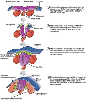
Neurulation is the process of formation of the neural plate and neural crests and their closure. It is completed at the end of the 4th week, when the posterior neuropore closes.
Neural plate and tube[edit | edit source]
The surface ectoderm above the notochord thickens and forms a longitudinally oriented formation of tall epithelial cells – the neural plate. At first, the extent of the neural plate corresponds to the cephalic process, later it exceeds its extent. Around day 18, the neural plate invaginates along the long axis and turns into a neural furrow extending into neural crests (distinct mainly in the cranial region – the beginning of brain development). At the end of the 3rd week, the bundles merge and turn into the neural tube - the basis of the CNS. The neural tube soon detaches from the surface ectoderm that closes over it – this is completed during the 4th week.
Formation of the neural crest[edit | edit source]
As the neural crests fuse, some of the ectoderm cells at the top of the crests become detached from their connections with other cells. They separate from both the neural tube and the ectoderm, travel dorsolaterally from the tube, and form the neural crest. Soon the bar divides into left and right, and both parts continue to travel around the circumference of the tube, giving rise to the sensory ganglia of the cranial and spinal nerves. Many neural crest cells travel in various directions and disperse in the mesenchyme.
- Structures originating from the neural crest
- Spinal ganglia and ganglia of the autonomic system,
- partly also ganglia n. V , VII , IX and X ,
- Schwann cells,
- cells of the brain membranes (mainly pia mater and arachnoid),
- pigment cells, elements of the adrenal medulla , part of the muscular and skeletal components of the head and teeth.
Clinical highlights[edit | edit source]
- Disorders of neurulation give rise to neural tube defects, which is one of the most common VVV.
- the most common and most severe defect is anencephaly or meroanencephaly (complete or partial absence of the brain).
Development of somites[edit | edit source]
During the formation of the notochord and the neural tube, the intraembryonic mesoderm proliferates on both sides, creating a strong longitudinal column - the paraxial mesoderm . It then passes laterally into the intermediate mesoderm , which further thins laterally into the lateral mesoderm . The lateral mesoderm is related to the extraembryonic mesoderm covering the yolk sac and amnion. Towards the end of the third week, the paraxial mesoderm begins to differentiate into paired cuboid formations - somites (primary segments). By the end of the fifth week, 42–44 pairs of somites are present. Somites contain a cavity in the middle called a myocoel, which soon disappears. The number of somites is a determining factor indicating the age of the embryo during the 4th and 5th week. The first somites appear in the occipital landscape, they rapidly increase in the caudal direction. Most of the axial skeleton develops from them, along with muscles and the skin dermis.
Development of the intraembryonic coelom[edit | edit source]
Intraembryonic coelom means the body cavity of the embryo. The basis of this cavity can be seen in small isolated coelomic cavities or sacs in the lateral and cardiogenic mesoderm. The sacs merge into a horseshoe-shaped cavity - the intraembryonic coelom. The cavity thereby splits the lateral mesoderm into two sheets:
- somatic (parietal sheet) – related to the extraembryonic mesoderm covering the amnion,
- splanchnic (visceral sheet) – passes into the extraembryonic mesoderm covering the yolk sac.
During the second month, the coelom divides into three cavities – pericardial, paired pleural and peritoneal.
Early development of the cardiovascular system[edit | edit source]
At the beginning of the third week, angiogenesis also occurs , which takes place in the extraembryonic mesoderm of the yolk sac, germinal shaft and chorion. Embryo vessels begin to form two days later. The reason why the vascular system develops so early is that there is little yolk in the yolk sac and there is an urgent need to transport nutrients to the growing embryo. At the end of the second week, nutrition is provided by maternal blood through diffusion through the embryonic coelom and yolk sac. During the third week, the primordial uteroplacental circulation develops.
Vasculogenesis and hematogenesis[edit | edit source]
Mesenchymal cells - angioblasts cluster and form isolated groups - blood islands . Small cavities appear inside the islets, which are created by the fusion of intercellular slits. Angioblasts flatten and transform into endothelial cells surrounding the sinuses. The spaces filled with endothelium soon merge to form a network of endothelial channels. Blood cells arise at the end of the 3rd week from endothelial elements (hemocytoblasts) in extraembryonic tissues. Hematopoiesis begins in the embryo itself only from the fifth week (mainly in the liver and spleen, bone marrow and lymph nodes). Fetal and definitive erythrocytes are probably derived from different precursors.
Primordial circulatory system[edit | edit source]
The heart and great vessels arise from mesenchymal cells in the cardiogenic zone. During the third week, paired endocardial heart tubes develop, which eventually merge into the primitive heart tube. The primary heart fuses with the vessels of the embryo, peduncle, chorion, and yolk sac to form the primordial circulatory system. At the end of the 3rd week, the blood is already circulating, the heart starts beating between the 21st and 22nd day. It is the first system to start working.
Further development of chorionic villi[edit | edit source]
Soon after their formation, the primary chorionic villi begin to branch. At the beginning of the third week, mesenchyme begins to grow into them, which begins to form their inner part - this creates secondary chorionic villi. The villi cover the entire surface of the chorionic sac. Vessels begin to develop in the mesenchyme, turning the villus into a tertiary villus . Cytotrophoblast cells proliferate and penetrate the syntitiotrophoblast to form a cytotrophoblastic sheath that surrounds the chorionic sac and attaches it to the endometrium. Villi that are attached via the cytotrophoblastic sheath are attachment villi.
Links[edit | edit source]
Related Articles[edit | edit source]
- Neurulation
- Prenatal development : Embryo • Fetus
- Gametogenesis • Fertilization • Types of eggs and their furrowing
- First week of embryo development • Second week of embryo development • Third week of embryo development • Fourth to eighth week of embryo development
Source[edit | edit source]
- MOORE, Keith L. – PERSAUD, T. V. N.. Zrození člověka. 1. edition. ISV, 2002. 564 pp. ISBN 80-85866-94-3.


