Muscle tissue


Muscle tissue has a unique cell architecture that allows for excitability and contraction. There are three main types of muscle tissue: skeletal, cardiac, and smooth, which are classified according to cell and tissue structure.
Skeletal (striated) muscle tissue[edit | edit source]
Skeletal muscle consists of muscle fibers, as well as connective tissue and blood vessels. The connective tissue encloses the muscle bundles and muscle fibers, and is also found at myotendinous junctions.
Schematic of the arrangement of connective tissue and muscle fibers.
Rhabdomyocytes[edit | edit source]
- diameter 10-100 μm; length 5-500 mm
- spindle-shaped
- multinucleated
- enclose myofibrils (diameter: 100-200 nm)
- linear arrangements of myofilaments in sarcomeres
- found along whole length of rhabdomyocyte
- desmin, plectin, nebulin, dystrophin stabilize myofibrils
- costamers attach myofibrils to sarcolemma via dystrophin-associated glycoprotein complex, which links the internal cytoskeleton to the ECM
- Duchenne muscular dystrophy: when the dystrophin protein is disfunctional, muscle contraction is impaired
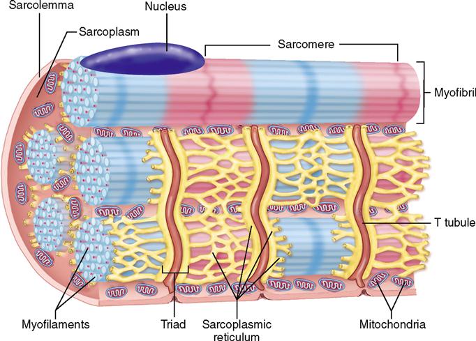
- sarcoplasm (equivalent of cytoplasm): has bulky, elongated mitochondria found between myofibrils
- myofilaments are of two types
- thin myofilaments (6-8 nm by 1 μm)
- G-actin form polymers of F-actin
- associated with tropomodulin, nebulin (stabilizes F-actin), dystrophin, titin (stabilizes myosin II --> elasticity), tropomyosin and troponin complex
- Troponin T (TnT): binds tropomyosin
- TnC: binds calcium
- TnI: inhibits interactions between actin and myosin
- attached to Z-line by α-actinin
- thick myofilaments (15 nm by 1.5 μm)
- complex of myosin heavy and light chains
- classification of muscle tissue depends on myosin heavy chain type
- associated with titin and MyBP-C protein
- attached to M-line by myomesin
- complex of myosin heavy and light chains
- thin myofilaments (6-8 nm by 1 μm)
- sarcoplasmic reticulum (equivalent of SER): wraps around myofibrils and forms terminal cisternae
- sarcolemma (equivalent of plasma membrane): forms tubular invaginations - T-tubules
- Triads consist of two terminal cisternae and one T-tubule in between
Schematic of muscle microstructure here.
See also https://is.muni.cz/auth/el/med/jaro2022/aVLBC0422s/um/011-Biochemistry_of_muscles_2022.pdf?lang=en
Sarcomere[edit | edit source]
The sarcomere (2-3 μm long, ~1 μm in contraction) is the structural and functional unit of myofibrils. It is a complex of thin and thick myofilaments between two adjacent Z-lines. Mnemonic: Zee Intelligent Animal Has Muscle.
- Z-line: dense attachment of positive ends of actin myofilaments between two adjacent sarcomeres
- I-band (isotropic): pale band having only thin myofilaments
- A-band (anisotropic): dense band where thin and thick myofilaments overlap
- H-zone: pale zone within A-band composed only of thick myofilaments
- M-line: dense site where thick myofilaments attach
During contraction, the Z-lines move closer to each other, and the I-band shortens.
Schematic of sarcomere here.
Neuromuscular junction (aka motor-end plate)[edit | edit source]
- axon terminal
- brings afferent signal from CNS
- contains numerous mitochondria and synaptic vesicles (acetylcholine, ACh)
- synaptic cleft (30 nm wide ): gap between axon terminal and post-synaptic membrane
- postsynaptic membrane
- non-myelinated terminal axonal branches interact with sarcolemma in junctional folds (shallow depressions)
- contains ACh-receptors and acetylcholinesterase
- action potential is delivered to sER via T-tubules
Proprioceptors[edit | edit source]
Sense changes in position
- muscle spindles
- found between muscle fascicles
- encapsulated by modified perimysium
- contain intrafusal fibers
- sensory axons wrap around intrafusal fibers but sense distension of extrafusal muscle fibers
- function in posture maintenance and opposing muscle group coordination
- Golgi tendon organs
- found at myotendinous junctions
- consist of encapsulated sensory axons penetrating through collagen bundles synapsing with inhibitory neurons
- sense tension; activate inhibitory neurons if it is excessive
Motor unit[edit | edit source]
- consists of a motor neuron and the muscle fibers that it innervates
- functions in coordination of muscle contraction
Connective tissue[edit | edit source]
- epimysium: dense irregular connective tissue with elastic fibers covering the entire muscle
- perimysium: loose connective tissue covering primary and secondary muscle bundles, together with vessels and nerves
- endomysium: reticular fibers and scattered fibroblasts covering muscle fibers and microvessels
Cardiac (striated) muscle tissue[edit | edit source]
- cardiomyocytes (15 μm diameter, 100 μm wide)
- cylindrical, branched cells
- 1-2 nuclei in the center of the cell
- myofibrils are also organized in sarcomeres
- sarcoplasmic reticulum contacts both sarcolemma and t-tubules
- T-tubules are deep and wide, located at the Z-line instead of overlap zone
- diads formed by just one sER cistern and T-tubule
- completely dependent on aerobic respiration, therefore there is an abundance of
- mitochondria
- myoglobin (to store oxygen)
- glycogen and lipid inclusions
- conductive cardiomyocytes: specialized cardiomyocytes in Purkinje fibers that conduct electrical stimuli, coordinating heart contraction
- myoendocrine cells: noticeable secretory granules (200-300 nm) that export atrial natriuretic peptides (for vasodilation and diuresis)
- intercalated discs: join cardiomyocytes, consist of
- adherent junctions
- desmosomes
- gap junctions
- interstitial connective tissue includes collagen and reticular fibers
- many capillaries and lymph vessels
Smooth muscle tissue[edit | edit source]
- smooth muscle cell (aka leiomyocyte: 20 μm long)
- fusiform shape
- centrally located nucleus
- involved in secretion, therefore, contain many mitochondria, RER and Golgi apparatus
- many pinocytic vesicles associated with SER and sarcolemma
- gap junctions present for the propagation
- maintains close contact with the ECM to allow for mechanotransduction (stimulation by pressure or stretch)
- can hypertrophy to 500 μm (pregnant uterus)

- contractile apparatus (oblique arrangement of microfilaments, not a sarcomere)
- thin filaments (associated with tropomyosin, caldesmon and calponin) and thick filaments (smooth muscle myosin) are present
- plasma membrane contains focal adhesions
- dense bodies contain α-actinin and serve to anchor thin filaments
- caveolae: invaginations of sarcolemma (50-70 nm) by which Ca2+ flows in (instead of T-tubules)
- intermediate filaments anchor to cytoplasmic dense bodies and attachment plaques of sarcolemma
- mechansm of contracton depends on myosin light chain kinase (MLCK) and calmodulin
- induction of contraction
- axon terminal and smooth muscle cell are separated by 10-20 μm (no junction)
- can be stimulated via
- signal from the CNS
- hormones (ex. epinephrine, ADH, oxytocin)
- mechanical stimulus
- spontaneously
- When Ca2+ is released, it binds to calmodulin, and the complex activates myosin light-chain kinase, which phosphorylates light chains and induces contraction
Mechanism of muscle contraction[edit | edit source]
Muscle contraction happens in several steps[1]:

- Neuromuscular transmission:
- Presynaptic action potential triggers opening of Ca2+ channels
- elevated Ca2+ causes ACh vesicles to move to the synaptic cleft and release their contents
- ACh diffusion binds to cholinergic receptors on the postsynaptic membrane and opens Na+ and K+ channels
- Botulotoxin inhibits release of ACh
- In myasthenia gravis, antibodies bind to cholinergic receptors, preventing contraction
- Curare poison (used on arrow tips) blocks ACh receptors
- Depolarization of postsynaptic membrane
- Action potential of the muscle fiber.
- Degradation of ACh by acetylcholinesterase.
- Connection between excitation and contraction:
- Action potential propagates through muscle fiber and stimulates the sarcoplasmic reticulum, which releases Ca2+
- Calcium binds to troponin C
- Troponin removes tropomyosin from the binding sites
- Contraction:
- Myosin head is in cocked state with ADP and Pi
- Myosin head binds to actin filament to form cross-bridge
- ADP and Pi are (separately) released during power stroke
- New ATP detaches myosin head from actin
- Phosphorylation of ATP re-cocks myosin head
- Cycle can repeat as long as there is ATP
- Relaxation:
- No more signal --> Ca2+ depletion
- Ca2+ unbinds from troponin C
- Active sites are covered by tropomyosin
Overview of Development[edit | edit source]
Satellite stem cells (from the mesoderm) differentiate into myoblasts, which differentiate into myocytes (these group together linearly), which further differentiate into myotubules (fused multi-nucleated myocytes).
References[edit | edit source]
Mescher, A. and Junqueira, L., 2018. Junqueira's basic histology. New York: McGraw-Hill, pp. 193-214.
Vaňhara, Petr et al. Guide To General Histology And Microscopic Anatomy. 1st ed., Masaryk University Press, 2020, pp. 4-5, 13.