Germ cells
Germ cells are cells that specialize in establishing a new individual. Mature egg and sperm carry only half the number of chromosomes, meaning they are haploid. Their fusion (penetration of sperm into the egg) creates the nucleus of a new diploid organism with the ability of very rapid cell division and differentiation.
Spermatogenesis[edit | edit source]
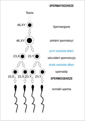
Spermatozoa arise in a process called spermatogenesis, which takes place in the seminiferous tubules in the testes, from puberty to death. Spermatozoa arise from primordial germ cells (spermatogonia) after a series of divisions ( mitotic and meiotic). Sertoli cells supply the necessary nutrients and hydrolytic enzymes needed to make mature sperm. Leydig cells, located in the interstitial space between the seminiferous tubules, produce testosterone. When sexual maturity is reached, primitive germ cells (spermatogonia) go through a number of divisions. The newly created elements follow one of the following two paths:
- may remain as undifferentiated cells - spermatogonia type A;
- differentiation during subsequent mitotic cycles to type B spermatogonia.
Type B spermatogonia give rise to primary spermatocytes, which enter the first meiotic prophase. At this point, the primary sperm contains 46 (44 + XY) chromosomes and 4N DNA (N = haploid set of chromosomes - 23 in humans - and the amount of DNA in this set). The first meiotic division results in secondary spermatocytes (22 + X or 22 + Y and the amount of 2N DNA). Secondary spermatocytes enter the second meiotic division very quickly and give rise to spermatids. There is no S-phase (i.e. no DNA synthesis) between the first and second meiotic divisions, so during the second division the amount of DNA is reduced by half (1N), creating elements with a haploid number of chromosomes and a quantity of DNA. This makes the sperm a highly specialized cell, carrying the genetic material of a male, equipped with great mobility, with the ability to swim upstream and penetrate its target - the female egg - at the time of fertilization.
Immobile sperm syndrome is caused by a lack of dynein or other flagella proteins. The disorder is usually associated with chronic inflammation of the respiratory tract, because the same effect affects the axonemas of the cilia of the airway epithelial cells ( Cartagener's syndrome).
Oogenesis[edit | edit source]
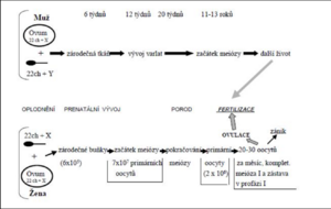
Oocytes develop in a process called oogenesis. It begins in the prenatal period, when in the ovaries embryo, all germ cells differentiate into primary oocytes. These enter into a meiotic division. However, the oocytes only enter the prophase of meiosis I and at this point the whole process stops until the onset of sexual maturity of the woman. During this prophase arrest, the yolk and proteins needed for embryo development accumulate and are stored early in fertilization. The egg thus gains in size.
During an individual's sexual maturity, several oocytes are stimulated periodically by hormones of menstrual cycle, leading to the completion of meiosis I. This results in an asymmetric division in which most of the yolk cytoplasm remains in the developed oocyte, now called secondary oocyte (ootida). The other half of the chromosomes are expelled from the oocyte, forming small residual structures - polar bodies. The process stops in metaphase II. The so-called cytostatic factor is responsible for this arrest.
The mechanism by which this happens is not yet fully understood. A signaling pathway involving the proto-oncogene c-mos (and its product) and MAP-kinase (mitogen-activated protein kinase) is thought to be involved. Mos-protein is also thought to be a protein kinase synthesized during oogenesis that activates MAP-kinase directly or indirectly. Activated MAP kinase stops oocyte development in metaphase II. During fertilization after sperm incorporation into the oocyte, the Mos-protein is degraded, the activity of the cytostatic factor is inhibited, so that the meiotic division is completed.
However, the second division is also asymmetric. A complete fertilized egg is formed. The reason for the asymmetric distribution during oogenesis is to ensure that there are enough nutrients in the ovine cytoplasm to withstand them until implantation in the uterus. At the onset of menarche, most of the 1-2 million fetal oocytes are degraded, leaving only 300,000-400,000; of these, only 350 to 400 will experience ovulation during a woman's reproductive age.
Germ cell differentiation[edit | edit source]
Testicular cell differentiation[edit | edit source]
The gonadal primordium is represented by a gonadal lamina, which is progressively colonized by extraembryonic primordial germ cells. The first recognizable event for testicular differentiation is evident from the 7th week of gestation, when primordial Sertoli cells begin to develop. Leydig cells begin to form at week 8 and proliferate by weeks 12-14. After birth they degenerate and temporarily disappear in the interstitial tissue, a new population does not begin to appear until the onset of puberty.
Ovarian cell differentiation[edit | edit source]
It starts later than the testes, but soon reaches a greater degree of maturity. In the 12th-13th week, some oogonia, located in the deepest layer of the cortex, enters a meiotic prophase. In the 7th month of gestation:
- all germ cells enter or stop in meiotic prophase;
- fetal granulosa cells produce estrogens (similar to the same stage of development, fetal testes produce testosterone);
However, the production of ovarian AMH (anti-Müllerian hormone) is demonstrable only after birth.
Capacity, fertilization and implantation processes[edit | edit source]
Fertilization is not just a fusion of sperm with an egg, but a combination of haploid male and female genomes. This fusion of the sperm with the egg takes place in the fallopian tube and the embryo is transferred by means of the fallopian tube cells to the uterine cavity, where it is implanted, ie the embryo is immersed in the uterine mucosa. During this transport, energy and nutrients for the embryo are obtained from the yolk cytoplasm. After implantation, there is a connection to the mother's blood circulation .
Sperm during movement of the female reproductive system undergoes so-called capacitance. Until this time, the sperm is not able to fertilize the egg. The mechanism of capacitance is not yet known in detail. It is likely that the female genital secretions alter the plasma membrane of the sperm so that it allows the sperm to bind to the egg. This binding takes place through the zona pellucida, an extracellular matrix rich in glycoproteins that surrounds the egg. It consists of three proteins: ZP-1, ZP-2, ZP-3 and ZP-3. ZP-3 serves as the actual receptor for sperm. This binding induces the release of hydrolytic enzymes from the acrosomal vesicles of the sperm head, which digest the pellucid zone at the junction with the sperm. The plasma membrane of the sperm and the egg fuse and the nucleus of the sperm with the genetic material is taken up by the egg. The fusion of the two membranes disrupts the cytoplasmic granule below the membrane, from which enzymes are released that digest the zona pellucida glycoproteins outside the membrane. This will then prevent the other sperm from joining the egg. At the same time, the "sleeping" cytoplasm of the oocyte is activated. The concentration of intracellular Ca 2+ increases, causing inactivation of the cytostatic factor with subsequent completion of meiosis, in which the maternal and paternal genomes stored in chromosomes fuse to form a nucleus of the zygote. This is followed by a very rapid division of embryonic cells.


