Paget's disease

Paget’s Disease (syn. morbus Paget, osteitis deformans, osteodystrophia deformans) is a bone disease characterized by a localized disorder of bone remodelling, in which there is excessive bone resorption followed by compensatory increased new formation of structurally incomplete bone. The newly formed bone is highly disorganized, we call it "felt bone". Bones changed in this way are more susceptible to fractures.
Affects either:
- one bone – monostotic form (15 %),
- more bones – polystotic form.
Predilection axial skeleton: spine, pelvis, femur, sacrum, skull.
Epidemiology[edit | edit source]
The occurrence of Paget's disease is common in Europe (Great Britain), North America, Australia, New Zealand, and is less common in Asia and Africa.
The disease typically begins in the 5th–6th century. decade in both men and women (male:female ratio 3:2).
Etiology[edit | edit source]
The causative cause is not yet known, genetic factors and viruses (environmental factors).
Pathogenesis[edit | edit source]
- Osteolytic phase: characterized by increased activity of abnormal osteoclasts (with several times more nuclei than normal osteoclasts).
- Mixed phase: increased osteolysis leads to increased new bone formation.
- Sclerotization phase: the new formation of bone tissue predominates.

Microscopic image of Paget’s disease ; Note the irregularity of the osteons

Microscopic image of healthy compact bone


Symptoms[edit | edit source]
70–80 % of patients asymptomatic. Others have:
- pain in the bones and adjacent joints, the pain is localized and dull, very often occurs at night
- bone deformities, skull, enlargement and deformities of vertebrae,
- headache,
- an increase in head circumference (the patient suddenly has a small hat),
- manifestations of nerve compression (hearing impairment)
- at the site of the newly formed bone, the formation of numerous arteriovenous connections, which lead to increased blood flow and a palpable increase in temperature above the affected area,
- bone fractures
Differential diagnosis[edit | edit source]
- osteomalacia,
- osteoporosis,
- osteoarthritis,
- bone metastasis,
- primary bone tumor,
- hyperparathyroidism.
Diagnostics[edit | edit source]
History: Positive family history in approx. 14 % of cases.
Imaging techniques: X-ray (lytic and sclerotic lesions), CT, radioisotope bone scan
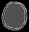
Skull in Paget’s Disease

Impairment of the right iliac blade


Laboratory diagnostics (markers of bone remodeling):
- bone resorption markers: deoxypyridinoline, N-telopeptide and C-telopeptide of collagen I (INTP, ICTP),
- neoplasia markers: ALP (bone isoenzyme),
- serum values of calcium, phosphorous and PTH: may be normal.
Complications[edit | edit source]
- bone: fracture, deformity, bone sarcoma,
- neurological: oppression of nerve roots, deafness
- increased blood flow to the bones can lead to hyper-circulation, with greater extent heart failure.
Therapy[edit | edit source]
- biphosphonates (alendronate, pamidronate, zoledrontate etc.),
- analogue of calcitonin,
- non-steroidal anti-inflammatory substances (NSPZL),
- supplementation of vitamin D and calcium,
- surgical therapy of bone deformities, fractures,
- regular checks.
Links[edit | edit source]
Related articles[edit | edit source]
- Disorders of calcium phosphate metabolism
- Indications of bone remodeling, markers of bone resorption
References[edit | edit source]
- Wikipedia. Paget´s disease [online]. [cit. 2009-10-06]. <en.wikipedia.org>.
- Wikipedia. Osteodystrophia deformans [online]. [cit. 2009-10-06]. <de.wikipedia.org>.
- GOLDMAN, L – AUSIELLO, D. Paget´s disease [online]. ©2007. [cit. 2009]. <MedlinePlus>.
- CARBONE, LD – DRIVER, K – LOHR, KM. Paget disease [online]. ©2008. [cit. 2009]. <emedicine.medscape.com>.