Hemorrhagic diatheses
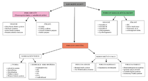
Hemorrhagic diatheses are bleeding conditions characterized by spontaneous bleeding manifestations or bleeding that is disproportionate to the underlying cause.
- Disorders of primary hemostasis (reaction of blood vessels at the site of injury + platelet activity):
- vasculopathy - unspecified vascular wall diseases;
- thrombocytopenia - reduced concentration of platelets;
- thrombocytopathy - disorder of platelet function.
- Disorders of secondary hemostasis (hemocoagulation) - coagulopathy.
Disorders of primary hemostasis[edit | edit source]
- They are clinically manifested by the formation of petechia, purpura, bleeding from the nose, gums, in the GIT, hematuria.
- Bleeding can be stopped with compression.
Vasculopathy[edit | edit source]
A. Congenital[edit | edit source]
- Teleangiectasia hereditaria haemorhagica (morbus Rendu-Osler-Weber)
AD is a disease in which microaneurysms form in the capillaries and veins of the skin, mucous membranes and internal organs (lungs, liver, brain, spleen).
- Marfan syndrome
AD is a congenital disease of the connective tissue, which is conditioned by a disorder in the formation of fibrillin (part of the amorphous component of the intercellular mass), patients live an average of 30 years, it affects several organ systems:
- skeleton – tall, slender figure, arachnodactyly, hyperflexibility of joints, dolichocephalia with prominence of tubera frontalia, pectus carinatum or excavatum, kyphoscoliosis;
- cardiovascular system - dissecting aneurysma of the aorta, there may be a redundant leaflet of the mitral valve;
- eyes – ectopia lentis (dislocation of the lens), myopia.
- Ehlers-Danlos syndrome
Congenital disease of the connective tissue, which is caused by a disorder in the formation of collagen, the most affected are:
- skin – extremely elastic, pulls out in long lashes, easily injured, even small traumas lead to wide-opening and poorly healing defects;
- joints – hyperflexibility (e.g. the thumb can touch the forearm during dorsiflexion);
- internal organs - the possibility of rupture of the wall of the large intestine, large arteries, hernias easily occur;
- eye – corneal rupture.
B. Obtained[edit | edit source]
Avitaminosis C ( scurvy)
Vitamin C is necessary for the synthesis of hydroxyproline and hydroxylysine, which are building blocks of collagen, vitamin C deficiency leads to bleeding manifestations (hemorrhagic gingivitis, subcutaneous and muscle hematomas) and to ossification disorders (poor fracture healing, in children Moller-Barlow disease)
Cushing's syndrome (hypercortisolism) and glucocorticoid therapy
An excess of glucocorticoids leads to a decrease in collagen synthesis.
- Some infections
Some infectious agents increase the permeability of the vascular wall with their toxins (inflammation - scarlatina - Streptococcus pyogenes, measles - morbilli - paramyxoviruses) or directly damage it (rickettsia - they parasitize inside endothelium)
- Henoch-Schönlein purpura
Immunopathological reaction III. type, when immune complexes (proven presence of IgA) are deposited in the vessel walls, especially in the skin and glomeruli (focal glomerulonephritis - hematuria), occurs mainly in children and adolescents, often after an infection of the respiratory system.
- Senile purpura
Increased fragility and loss of elasticity of blood vessels in old age.
Thrombocytopenia[edit | edit source]
Decrease in platelet concentration (normal value is 150-300×109/l[1]), příčiny:
- Nedostatečná tvorba – aplastické a hypoplastické syndromy, porucha megakaryocyto- a trombocytopoézy;
- Nadměrná destrukce nebo konsumpce – autoprotilátky proti trombocytům, TTP, ITP, DIC, HUS, umělé srdeční chlopně, Wiskottův-Aldrichův syndrom
- Sekvestrace destiček ve slezině – hypersplenismus při splenomegalii
- Wiskott-Aldrich syndrome
GR disease, consisting in a disorder of the membrane glycoprotein on the surface of T-cells (primary immunodeficiency) and platelets (increased destruction in the spleen - thrombocytopenia), the disease is characterized by repeated infections, bleeding and eczema.
- Thrombotic thrombocytopenic purpura (TTP)
- Bleeding (purpura) from thrombocytopenia (thrombocytopenic) caused by the consumption of platelets in the formation of thrombi (thrombotic).
- The cause is apparently damage to the endothelium with the release of the von Willebrand factor causing platelet aggregation with simultaneously reduced activity of the protease that cleaves the von Willebrand factor (either autoantibodies against this protease or its congenital defect) - platelet microthrombi are formed without consumption of coagulation factors - the consequence is ischemia (e.g. . neurological symptoms and renal failure), bleeding and microangiopathic hemolytic anemia (breakdown of erythrocytes by thrombi).
- Idiopathic Thrombocytopenic Purpura (ITP)
The formation of autoantibodies against platelets, which are then phagocytosed in the spleen (possible therapy is splenectomy), can occur together with SLE. Antibodies can be:
- complexes of viral antigen with antiviral IgG that bind to platelets;
- anti-viral antibodies cross-reacting with platelets;
- autoantibodies against platelet membrane surface proteins.
- ITP can therefore arise acutely after a viral infection (ad 1. and 2.) or it can be chronic (ad 3.).
- Disseminated intravascular coagulation (DIC)
It is caused by the pathological occurrence of tissue factor in the blood (generalized blood clotting occurs and thus consumption of plasma coagulation factors - consumptive coagulopathy, which is the cause of subsequent bleeding).
- Possibilities of penetration of tissue factor into the vascular bed:
- extravascular cells – they enter the blood e.g. during childbirth, during trauma, operations, penetration of tumor cells into the circulation;
- pathological blood cells – in myelo- and lymphoproliferative diseases;
- expression in the membrane of activated endothelium and monocytes – activation by endotoxin, systemic inflammation;
- release from hemolyzed erythrocytes.
- Pathogenesis:
- Tissue factor activates factor VII (external pathway) and this triggers the coagulation cascade leading to the formation of fibrin - hemocoagulation becomes generalized with the formation of microthrombi in various organs - these clog the vessels and, in addition, mechanically damage platelets - the result is the consumption of coagulation factors (coagulopathy) and thrombocytopenia – i.e. a combined primary and secondary hemostasis defect that leads to bleeding.
- Bleeding occurs from surgical wounds, punctures, from the gums (and elsewhere in the GIT), hematuria, epistaxis, hematomas are formed, bleeding into internal organs can occur, incl. brain, microthrombi and microemboli in the periphery can lead to pre-gangrenous changes (acrocyanosis) and signs of organ disorders (liver, kidneys, lungs).
- At the same time as increased coagulation, fibrinolysis is also activated, which is manifested by an increase in FDP (including D-dimer) and a decrease in fibrinogen (clinical experience is that the risk of bleeding and its degree roughly correspond to a decrease in fibrinogen concentration).
- Hemolytic uremic syndrome (HUS)
- A disease mainly of preschool children characterized by a combination of acute renal failure with oliguria and microangiopathic hemolytic anemia with thrombocytopenia, in addition hemolytic icterus.
- The basis of the syndrome is damage to the endothelium in the glomeruli, vas afferens and smaller arteries. Toxins of some bacteria (e.g. enterohemorrhagic verotoxin E. coli or shigatoxin Shigella dysenteriae, as well as endotoxin of a number of bacterial strains) and viruses (e.g. from the group [[Coxsackie]) are used etiologically ]), the formation of fibrin and aggregation of platelets are deposited on the damaged endothelium - hyaline thrombi are formed clogging the capillaries of glomeruli and afferent arterioles - ischemic damage to the cortex occurs (up to necrosis, but usually it is milder - dispersion necrosis of individual tubules, dilation and reduction of the lining of proximal tubules) , which results in uremia, the hemolysis is extracorpuscular, by breaking the erythrocytes against the fibrin fibers in the capillaries (similar to DIC).
Thrombocytopathy[edit | edit source]
A. Congenital[edit | edit source]
- Disorders of platelet adhesion and aggregation
- These include disorders of surface glycoproteins gpIb-IX (Bernard-Soulier syndrome) and gpIIa-gpIIIb (Glanzmann thrombasthenia) and decreased von Willebrand factor (von Willebrand disease).
- Disorders of platelet secretion
- This includes Heřmanský-Pudlákand Chediak-Higashi - platelet granule defect.
B. Obtained[edit | edit source]
This mainly includes the irreversible blockade of platelet cyclooxygenase (synthesis of TxA2, which is important in the aggregation and secretion of platelets) acetylsalicylic acid.
Disorders of secondary hemostasis (coagulopathy)[edit | edit source]
- Clinical manifestations are similar to disorders of primary hemostasis (epistaxis, bleeding from the gums, into the GIT, hematuria, menorrhoea), but petechia and purpura are absent and there is bleeding into deep tissues (joints, retroperitoneum, brain) and poor wound healing.
- Compression does not stop the bleeding, it usually continues in the form of long-term blood seepage.
A. Congenital[edit | edit source]
- These are often GR diseases linked to the X-chromosome (mainly affects men, less women), only one coagulation factor is affected.
Hemophilia A
- Factor VIII deficiency (synthesized in the liver, also contained in erythrocyte granules, circulates freely in the plasma, binds to the von Willebrand factor formed in the endothelium and is thereby stabilized), is clinically serious only when factor VIII drops below 1% of the normal concentration, then there is unstoppable bleeding after trauma and operations, typical is 'spontaneous bleeding into the joints.;Hemophilia B
- Factor IX deficiency.
- Hemophilia C
- Factor XI deficiency.
- Afibrinogenemia
- No fibrinogen is present in plasma.;Dysfibrinogenemia
- The presence of a defective form of fibrinogen (reduced blood coagulation, but some mutations can, on the contrary, cause an increased tendency to convert fibrinogen to fibrin - formation of thrombosis).
B. Obtained[edit | edit source]
- Several coagulation factors are affected at the same time, the main causes are:
- Hepatic insufficiency
- The liver is the source of the coagulation factors of the so-called prothrombin complex (which are characterized by the carboxylation of some glutamic acid residues dependent on vitamin K) – factors II, VII, IX, X, proteins C and S, other proteins produced in the liver are factor I, V, XI, XII, and XIII; the liver is also a place of storage and metabolic activation of vitamin K. If its disease is accompanied by splenomegaly, it can indirectly cause thrombocytopenia, plasmin (fibrinolytic agent) is also inactivated in the liver, in their disease it is activated [ [fibrinolysis]].
- Vitamin K deficiency
- Vitamin K participates in γ-carboxylation of glutamic acid residues of factors II, VII, IX, X and proteins C and S in the liver. γ-carboxylation is essential for their adhesion to phospholipid surfaces. The cause of vitamin K deficiency is its insufficient resorption in the intestine.
- Therapeutic administration of anticoagulants
- E.g. administration of warfarin, which blocks the action of vitamin K.
- DIC - see above.
Links[edit | edit source]
Related Articles[edit | edit source]
Source[edit | edit source]
- POVÝŠIL, Ctibor – ŠTEINER, Ivo – BARTONÍČEK, Jan, et al. Speciální patologie. 2. edition. Praha : Galén, 2007. 430 pp. ISBN 978-807262-494-2.
References[edit | edit source]
- ↑ POVÝŠIL, Ctibor – ŠTEINER, Ivo – BARTONÍČEK, Jan, et al. Speciální patologie. 2. edition. Praha : Galén, 2007. 430 pp. pp. 68. ISBN 978-807262-494-2.

