Hemocoagulation
Hemocoagulation is one of processes leading to termination of bleeding (hemostasis). The main principle is the formation of a fibrin network that captures erythrocytes, leukocytes and platelets from the bloodstream and forms a definitive thrombus, replacing the primary (white) thrombus. This process is controlled by a number of coagulation factors. The exact sequence of events leading to hemocoagulation is called the coagulation cascade.
Stages of hemocoagulation
Haemocoagulation consists of the following phases:
- Formation of prothrombin activator from factor X and V
- Conversion of prothrombin to thrombin
- Conversion of fibrinogen to fibrin
Formation of prothrombin activator
The presence of the enzyme thrombin, which is formed from prothrombin, is the key to the conversion of fibrinogen to fibrin. Thus, the formation of prothrombin activator is the limiting factor in the whole process. Prothrombin activator is produced by an extrinsic or intrinsic hemocoagulation cascade.
Extrinsic hemocoagulation cascade
Damage to the blood vessel wall results in the release of tissue thromboplastin (factor III) into the blood. Contact with tissue factors activates coagulation factor VIIa, which in turn activates factor X in the presence of Ca2+ ions. The latter binds to tissue factor phospholipids and, with the help of factor V, forms prothrombin activator. In the presence of Ca2+ and platelet phospholipids, it converts prothrombin to thrombin. Thrombin activates other factor V molecules (an example of positive feedback).
Intrinsic hemocoagulation cascade
When contact occurs between blood and a negatively charged or wetting surface, factor XII activation occurs. By its subsequent reaction with prekalikerin and high molecular weight kininogen, factor XI is converted to its active form. Factor IX is then activated in the presence of Ca2+. In the presence of factors VIIIa and IXa, platelet phospholipids and calcium ions, factor X is activated. This, together with factor Va, forms prothrombin activator, which is involved in the conversion of prothrombin to thrombin. Factors V and VIII are activated by thrombin in a positive feedback loop.
Conversion of prothrombin to thrombin
Prothrombin (factor II) is a plasma protein produced in the liver. Its production is strongly dependent on vitamin K. It is continuously leaked into the bloodstream and is not stored (plasma concentration is 150 mg/l)[1]. It is modified by prothrombin activator in the presence of Ca2+ ions (see above).
Conversion of fibrinogen to fibrin
Fibrinogen (factor I) is a plasma protein formed in the liver that belongs to the β-2-globulins. The catalytic action of thrombin results in the cleavage of several peptides to form monomeric fibrin, which polymerizes to form a fibrin net. This is initially loose and has to be stabilised. This is ensured by the activated fibrin stabilising factor (factor XIII) with the participation of Ca2+ by covalent linking the individual chains.
Coagulation factors
Coagulation cascade:
Coagulation factors are proteins that circulate in plasma in an inactive state. Their main function is to enable hemocoagulation (blood clotting). Most of them are produced by the liver.
| Factor | Name
Alternate Name |
Function |
|---|---|---|
| I | fibrinogen | cleavage of several peptides produces monomeric fibrin, which further forms a fibrin network |
| II* | protrombin | its active form (IIa) activates factors I, V, VII, VIII, XI, XIII, protein C and platelets |
| III | tissue thromboplastin
"tissue factor" |
factor VIIa cofactor |
| IV | Ca2+ | binding of coagulation factors to phospholipids |
| V | proaccelerin, labile factor, accelerating globulin | factor X cofactor – ensure the conversion of prothrombin to active thrombin |
| VI | older name for factor Va | – |
| VII* | proconvertin | activates factors IX, X |
| VIII | antihemophilic factor (AHF)
antihemophilic factor A – antihemophilic globulin (AHG) |
factor IX cofactor |
| IX* | The Christmas Factor
plasma thromboplastic component (PTC) - antihemophilic factor B |
activates factor X |
| X* | Stuart-Prower factor** | activates factor II |
| XI | plasma thromboplastin precursor
plasma thromboplastin antecedent (PTA) - antihemophilic factor C |
activates factor IX |
| XII | The Hageman factor
glass factor |
activates factor XI, VII and prekallikrein |
| XIII | fibrin stabilizing factor
The Laki-Lorand Factor |
|
| von Willebrand factor | binds to factor VIII, enables platelet adhesion | |
| high molecular weight kininogen (HMWK)
The Fitzgerald Factor |
supports the mutual activation of XII, XI and prekallikrein | |
| prekallikrein (PKK)
The Fletcher Factor |
activates factor XII and prekallikrein, cleaves HMWK | |
| kallikrein | ||
| platelet phospholipids |
* vitamin K dependent
** named after the first two patients (Mr R. Stuart and Miss A. Prower) in whom factor X deficiency was described
Anticlotting mechanisms
The modulation of the response that maintains the smooth flow of blood in the blood vessels is called the fluid-coagulation balance. The inhibitory system consists of three parts:
- Blood flow, which washes away and dillutes coagulation factors
- Intact vascular endothelium provides a non-wetting surface and prevents contact with interstitial, negatively charged connective tissue
- Humoral inhibition is the most important and precise system of regulation and includes antithrombin III, heparin and protein C
- Antithrombin (also antithrombin III, ATIII) binds to thrombin and other coagulation factors and inhibits them (this effect is significantly amplified by heparin).
- Thrombomodulin together with thrombin (negative feedback) activates protein C and protein S, which cleaves coagulation factors.
Protein C and protein S are also vitamin K dependent.
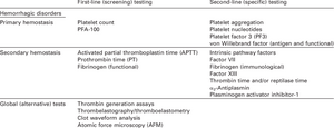
Examination of hemocoagulation
Blood clot removal
When the blood thrombus has served its function, it must be removed. This is done in two steps. First, the thrombus is retracted by contraction of the actin and myosin filaments of the platelets. This reduces their volume and allows the damaged tissue to regenerate. The next step is fibrinolysis. It is a process in which, with the help of an enzyme plasminogen the fibrin network will dissolve. Tissue plasminogen activator converts plasminogen to plasmin, which subsequently dissolves fibrin fibers and factors V, VIII, XII. The plasminogen system maintains microcirculation by dissolving clots in capillaries.
Targeted influencing of haemocoagulation
Decreasing coagulation
The decrease in coagulation is intentionally induced:
- in coagulation system ilnesses (e. g. some genetic disorders);
- when reducing the speed of blood flow through certain parts of the body (e.g. prevention of thromboembolic disease of the lower limbs before surgical procedures, atrial fibrillation);
- when blood comes into contact with artificial materials (e.g. hemodialysis, extracorporal circulation).
Anticoagulants are used, most often heparin and its derivates (parenterally) – supports anticlogging mechanisms and warfarin (p.o.) – inhibits vitamin K.
In vitro we use anticlogging agents if we don't want the blood in the tube to clot. They usually work on the principle of Ca2+ ion binding (clotting can therefore be restored by re-introducing calcium ions).
Increasing coagulation
Coagulation increase is desired in coagulation factors deficits (e.g. in haemophilia), when missing factors or plasma is applied.
Pathology
- Fibrinogen is one of non-specific inflammation markers.
- Since coagulation factors are synthesized in the liver, coagulation parameters are a sensitive indicator of liver injury.
- Increased tendency to blood clotting can cause thromboses and embolisms.
- Deficiency of certain coagulation factors can lead to bleeding manifestations (e.g. hereditary haemophilia).
- Some conditions can lead to combined disorders, thrombs are being formed and due to the consumption of coagulation factors, severe bleeding occurs. Such a concerning complication is disseminated intravascular coagulation (DIC).
Links
Related articles
- Hemocoagulation versus anticoagulation
- Coagulation versus aglutination
- Blood draws for testing
- Blood count
- Blood clotting test
- Bleeding disorders test
- Erythrocyte sedimentation rate
- Biochemical analysis of blood
- Laboratory acid-base balance test
- Hemoculture
- CRP
- PCT
External links
- Mechanisms in Medicine: The Coagulation Cascade(video)
- Hemokoagulace (czech wikipedia)
- Atlas fyziologie a patofyziologie – hemostasis
References
- ↑ KITTNAR, Otomar – ET AL.,. Lékařská fyziologie. 1. edition. Grada, 2011. 790 pp. ISBN 978-80-247-3068-4.
Used literature
- GANONG, William F. Přehled lékařské fyziologie. 20. edition. Galén, 2005. pp. 546–549. ISBN 80-7262-311-7.
- KOOLMAN, J – ROEHM, KH. Color Atlas of Biochemistry. 2. edition. Thieme, 2005. pp. 290–291. ISBN 1-58890-247-1.
- KITTNAR, Otomar. Lékařská fyziologie. 1. edition. Grada, 2011. 790 pp. ISBN 978-80-247-3068-4.


