Embolism
Definition: the introduction of a moving object (inset - embolus) into the place of the vascular bed, where the narrowing prevents its further movement (consequence: arteries - ischemia, veins - venostasis).
Embolus:
- thrombus – thromboembolism (thrombotic embolism);
- fat - fat embolism;
- cells (tumor, amniotic fluid embolization) – cellular embolism;
- air – air embolism;
- DNA, atheroma masses – subcellular embolism;
- foreign body – e.g. broken catheter...
| where from (what) | where/to |
|---|---|
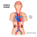
| lower limb veins (thrombus) | pulmonary arteries |
| right heart (thrombus) | pulmonary arteries |
| jugular veins (air) | pulmonary arteries |
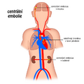
| left heart (thrombus) | arteries of the brain, kidneys, spleen, mesenteric arteries, DK arteries |
| aorta (thrombus, atheroma masses) | as in the left heart |
| pulmonary veins (air) | as in the left heart |
Successive embolism - repeated embolism.
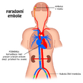
Paradoxical embolism - an embolus (thrombus) enters the arterial circulation from the peripheral veins through the through foramen ovale (the condition is that the pressure in the right atrium of the heart is greater than in the left atrium - e.g. with simultaneous embolization of the pulmonary artery or hypertrophy of the right heart), or from aorta to the pulmonary artery through the ductus arteriosus.
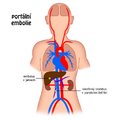
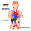
Retrograde embolism – movement of the thrombus against the blood flow (e.g. from the IVC to the hepatic veins with increased intrathoracic pressure – coughing, turning on the abdominal press...).
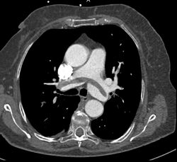
- The embolus usually fills the entire lumen of the vessel, it can also attach to the bifurcation of the vessels (straddle embolus).
- The result of embolism is reflex vasoconstriction of the blocked vessel.
Thrombembolism[edit | edit source]
Typical blockage of the pulmonary artery occurs when a thrombus is torn from the veins of the lower limbs (massive embolization leads to reflex cardiac arrest and death, survivors develop a hemorrhagic heart attack).
Fat embolism[edit | edit source]
In case of bone injury (bone fragment, vein injury), blunt injury of subcutaneous and fatty tissue, burns. It is recognizable only microscopically (droplets of fat in the capillaries, they can push into the general circulation and we find them in the capillaries of the brain and glomeruli of the kidneys).
Cellular embolism[edit | edit source]
The spread of tumor metastases or embolism of amniotic fluid - suction into the uterine veins during childbirth and embolism into the pulmonary capillaries, where we find the components of the amniotic fluid - vernix (smear = peeled off fetal epithelia, hairs, parts of meconium) - leads to the formation of DIC, because in the cell membrane tissue thromboplastin (factor III) is present in the fetus and there is a risk of developing hemorrhagic shock and exsanguination of the mother.
Air embolism[edit | edit source]
If air enters the veins (peripheral veins during operations on the thyroid gland or posterior cranial fossa), it enters the right heart and pulmonary arterioles - death when 100-200 ml is sucked in, or enters the pulmonary veins - it enters the left heart and brain - even a small amount of air will cause ischemia and death (0.5-3 ml) + caisson disease (N 2 bubbles during decompression, especially in fat-rich tissues - CNS).
Subcellular embolism[edit | edit source]
DNA from disintegrated cells - after chemotherapy, atheroma mass from a ruptured plaque (especially from the aorta to the renal arteries) is injected.
Embolism schemes[edit | edit source]
Links[edit | edit source]
Related articles[edit | edit source]
External links[edit | edit source]
Source[edit | edit source]
- PASTOR, Jan. Langenbeck's medical web page [online]. [cit. 2009]. <https://langenbeck.webs.com/>.


