Endocytosis
Endocytosis is an energy-demanding and material-demanding process characteristic of animal cells, during which both molecules and other cells from the external environment are absorbed by them. The opposite process, where the cell gets rid of substances by releasing them into the environment, is exocytosis. Cells are separated from the external environment by a cytoplasmic membrane. Some hormones, lipoprotein particles, viruses, antibodies, but also damaged cells or bacteria get into them through endocytosis.
The endocytosis process itself can take place in two basic ways – across the membrane or in the membrane continuum.
Through the membrane[edit | edit source]
In this way, some ions and molecules in particular are transported into the cell, which can penetrate the membrane passively in the direction of the osmotic, concentration or potential gradient. This includes simple (facilitated) diffusion and active transport.
In the membrane continuum[edit | edit source]
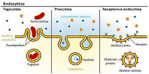
It is generally characteristic of this mechanism that substances absorbed by the cell remain separated from the surrounding cytoplasm by a biological membrane. The absorbed content is present inside the cell in the form of a cytoplasmic vesicle - an endosome. The category of endosomes includes pinosomes (formed during pinocytosis), phagosomes (during phagocytosis) and receptosomes. On average, these vesicles range in size from 0.3–1 µm. Smaller vesicles are located on the periphery of the cytoplasm, larger ones are usually located deeper. An important feature of endosomes is the acidity of their internal environment, caused by the accumulation of proton pumps. The pH in them ranges from 5 to 6. The acidic environment on the one hand changes the content of endosomes, on the other hand it participates in the transport of Fe2+ ions into the cell – this transport is mediated by the glycoprotein transferrin. Endosomal vesicles most often transport their contents to lysosomal transport vesicles, i.e. primary lysosomes (organelles involved in the degradation of already unwanted material for the cell), thereby creating lysosomes in the narrower sense of the word, i.e. secondary lysosomes. Lysosomes contain hydrolytic enzymes that degrade the contents of endosomes into substances that can be further used by the cell.
Pinocytosis[edit | edit source]
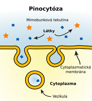
Pinocytosis occurs almost constantly in most animal cells. Cells absorb droplets of extracellular fluid and substances dissolved in it.
Phagocytosis[edit | edit source]
This is a process in which the cell actively creates spikes in order to absorb larger solid particles - it is used by all cells of the body, since most of the substances important to the cell are large polar molecules that cannot spontaneously penetrate through the cell membrane. In addition to "simple molecules", this process can also absorb bacteria and other microorganisms that have penetrated the organism - in this case we are talking about a special subtype of phagocytosis, heterophagy (cells capable of heterophagy include, among others, some types of leukocytes or some protozoa - amoebas). Autophagy is the process in which the cell absorbs the organism's own or damaged parts.
Receptor-mediated endocytosis[edit | edit source]
Substances are taken up only after they bind to specific receptors. These receptors are glycoproteins that have the ability to specifically bind substances (ligands) to be absorbed. They are located mainly on the surface of the cytoplasmic membrane, where endocytotic invagination also begins. At the base of the pit (emerging invagination) we find a layer of proteins, of which clathrin plays the biggest role in the endocytosis process. Its molecules have the shape of a tripod - a triskelion. Clathrin molecules accumulate under the section of the membrane where the receptor and ligand bind and their polymerization occurs. Spatial networks are formed around the bladder, thanks to which the receptor pit (coated pit) and later the vesicle itself (coated vesicle) are formed. The clathrin network then gradually depolymerizes. Vesicles that contain receptor-bound ligand (this binding is strongest at pH around 7) fuse with other small vesicles to form the early endosome. Here the pH is already lower (5–6), mediated by proton pumps. At lower pH, the binding of ligand and receptor is gradually loosened. The membrane and receptors that were part of the vesicle are not degraded in the lysosomal apparatus, but are recycled, returning to the plasma membrane.
Links[edit | edit source]
Related Articles[edit | edit source]
Source[edit | edit source]
- VAJNER, Ludek, et al. Medical histology I : General Cytology. 1. edition. 2010. 0 pp. ISBN 978-80-246-1860-9.
- MESCHER, Anthony L. Junqueira's basic histology: text and atlas. Thirteenth edition. New York [etc.]: McGraw-Hill Medical, 2013. ISBN 978-0-07-178033-9.


