Congenital developmental defects of the newborn requiring an urgent solution
Major urgent congenital developmental defects (CDD) include:
- congenital diaphragmatic hernia
- esophageal atresia
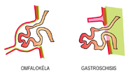
- abdominal wall defects (omphalocele and gastroschisis)
- neural tube clefts
- critical congenital heart defects and others.
Diaphragmatic hernia[edit | edit source]
The prevalence of diaphragmatic hernia is 1:2000–4000. Mortality is 25.9%. Up to 80% of all congenital lung anomalies originate from diaphragmatic hernias. In 95% of cases, the hernia is left-sided, the so-called Bochdale hernia, and is located lumbosacral on the left [1], in the remaining 5% it is right-sided, the so-called Morgagni hernia, and with sternocostal to the right. [1] A diaphragmatic hernia is not covered by a hernial sac, so it is called a false hernia. A defect in the diaphragm may be small or it may be completely absent. A hernia can include part of the Small Intestine and Large Intestine, Stomach, sometimes Liver, Spleen or even Kidney.
Clinical picture
Severe Respiratory distress syndrome develops immediately after birth (dyspnea, cyanosis, tachycardia), dyspnea subsides in an elevated position. The heart moves to the healthy side, so the location of the echo can change, which is called migration of echoes, the so-called Peter's sign. Breathing is usually weak, on the side of the hernia there is a drumbeat on the chest. Twisting can be heard on the chest, but the abdomen is sunken. The first critical 72 hours after birth are important for survival. After this period the disease manifests itself in chronic respiratory and GIT difficulties.
Shortness of breath, cyanosis, dextrocardia are listed as 'diagnosis. A native x-ray will confirm the diagnosis. The air filling of the intestines is missing in the abdomen, the mediastinum is displaced, in the left part of the chest you can see circular clearings reminiscent of loops of intestines, which have the shape of a signet ring. Prenatal diagnosis is also possible - ultrasound, polyhydramnios.
Therapy: intubation before surgery, gastric tube to suck out stomach contents (prevention of aspiration). We position the patient on the affected side and raise his upper body. Inhalation of surfactant, inhalation of NO (causes vasodilation - reduction of pulmonary hypertension). Then surgical reposition of the organs into the abdominal cavity. Covering a diaphragmatic defect is an urgent and life-saving procedure.
Esophageal atresia[edit | edit source]
The prevalence of esophageal atresia is 1:3000–4000. Mortality is 17%. A tracheoesophageal fistula appears in 85% of patients, and other anomalies in up to 50% of patients. Esophageal atresias are classified according to Vogt - the most common type is the so-called Vogt 3B (86%) - the finding of a lower esophagotracheal fistula with an upper cecum. VACTERL syndrome (Vertebral defect, Anal atresia, Congenital heart disease, Tracheo-esophageal fistula, Renal + radial limb defect)
Symptoms include polyhydramnios, salivation, coughing episodes, retching, cyanosis and abdominal distension. An x-ray with aqueous contrast is performed as proof. Treatment is performed operatively by closing the tracheoesophageal fistula as soon as possible after delivery, because otherwise there is a risk of aspiration[1], for surgery the patient must be of a certain weight, premature infants are indicated for parenteral nutrition, and then comes surgery.
Atresia and critical duodenal stenosis[edit | edit source]
The prevalence of duodenal atresia and stenosis is 1:6000. Mortality is 4.8%. In 70% other anomalies also occur, in 30% it is associated with Down syndrome. Among the symptoms' are polyhydramnios, vomiting in the first 24 hours, in 90% of the vomits contain a biliary admixture. The disease is demonstrated prenatally by US, postnatally by native x-ray in the suspension, which shows a typical image of two levels[1]. Surgical Therapy - duodenoduodenal anastomosis.[1]
Atresia and stenoses of the jejunum and ileum[edit | edit source]
The prevalence of atresias and stenoses of the jejunum and ileum is 1:1000–10,000. Mortality is around 3.8%. Clinical picture: prenatally – polyhydramnios, postnatally – in the first 36 hours, the development of a clinical picture of moderate ileus. Vomiting with an admixture of bile occurs, the abdomen is distended and shortness of breath appears due to the high state of the diaphragm, meconium does not pass, dehydration with hypochloremia develops and the patient loses weight.[1]
There are four types of atresia:
- type I – simple atresia, membrane in continuity with intestinal wall
- type II – simple atresia, discontinuity of the membrane with the intestinal wall, reduction of the length of the intestine
- type III – multiple atresia
- type IV – "apple peel" deformity, missing mesentery, reduction of the total length of the intestine
They are diagnosed prenatally. Dilation of the intestinal loops is visible on the US, and a native X-ray of the abdomen in the hanging position is performed postnatally. Atresia or stenoses are removed surgically. An atretic or stenotic segment of bowel is removed and an end-to-end anastomosis is performed.[1]
Atresia of the anus and rectum[edit | edit source]
Anal and rectal atresia are congenital closures or narrowing of the distal part of the intestine, which arise as a result of failure to separate the lower intestine from the ventrally located urogenital system during embryonic development. They are often associated with other congenital developmental defects (CDD) such as esophageal atresia, CDD of the urogenital system, lumbar and sacral spine.
The incidence is 1:1500[1] and 2 forms are distinguished:
- high atresia' - the blind end is above the levator ani muscle (40% of cases);
- low atresia - the blind end is below the levator ani muscle (60% of cases).[1]
Defects in the abdominal wall[edit | edit source]
The prevalence of abdominal wall defects is 1:6000–8000. We distinguish gastroschisis, which has a mortality rate of 17.6% and is a defect of the abdominal wall, usually to the right of the navel. There is eventration of uncovered intra-abdominal organs. Another defect is omphalocele, which has a mortality rate of 22.2% and is a herniation of intra-abdominal organs into the hernia sac. This hernia is originally a physiological hernia during the development of the midgut, which did not return to the abdominal cavity as a result of a developmental defect. Other anomalies also occur in 50% of patients, often chromosomal aberrations. Inferior midline syndrome' includes vesicointestinal fissure, anal atresia, colonic agenesis, bladder exstrophy, and upper midline syndrome includes congenital heart malformations, diaphragmatic and sternal defects.
!! transport of a newborn with a congenital GIT defect is necessary with an open gastric tube in place !!
Critical Congenital Heart Defects[edit | edit source]
Critical congenital heart defects are those that require acute intensive care. The prevalence of these defects is 2-3/1000, which is around 19% of newborns with a birth weight below 2500 g. Premature infants mainly have ductus arteriosus patens (PDA), coarctation of the aorta (COA), ventricular septal defect (VSD). In full-term infants, it is transposition of the great arteries (TGA).
The manifestation of critical congenital heart defects (CHD) is cyanosis and heart failure.
- hypoxemia → pulmonary vasoconstriction → increase in pulmonary vascular resistance → decrease in pulmonary flow → increase in R-L shunts, FOA, PDA → increase in venous admixture → hypoxemia, … (circulus vitiosus)
Cyanotic critical CHD[edit | edit source]
- 5 T: truncus arteriosus, tricuspid atresia (stenosis, pulmonary atresia), tetralogy of Fallot, transposition of the great arteries (TGA), total anomalous pulmonary venous return
The most common critical VSV is TGA, which is 50% isolated and 50% associated with another defect.
Heart defects where the main manifestation is heart failure[edit | edit source]
- COA (coarctation of the aorta), IAA (interruption of the aortic arch), AS (aortic stenosis), TGA (transposition of the great vessels), VSD (significant ventricular septal defect) , PDA (Ductus arteriosus patens)
Duct dependent CHD[edit | edit source]
- PS/PD, AS, COA, IAA
In ductus-dependent developing cardiac vasculature, it is important to leave the ductus open or to reopen it. Therapy: PGE 1 0.01-0.05 µg/kg/min i.v. continuously (Alprostan); AE: hypoventilation, apnea, febrile, diarrhea
Links[edit | edit source]
Related Articles[edit | edit source]
References[edit | edit source]
Sources[edit | edit source]
- BENEŠ, Jiří. Studijní materiály [online]. [cit. 2010]. <http://jirben.wz.cz>.
Literature[edit | edit source]
- HRODEK, Otto – VAVŘINEC, Jan, et al. Pediatrie. 1. edition. Galén, 2002. ISBN 80-7262-178-5.
- ŠAŠINKA, Miroslav – ŠAGÁT, Tibor – KOVÁCS, László, et al. Pediatria. 2. edition. Bratislava : Herba, 2007. ISBN 978-80-89171-49-1.

