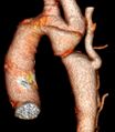
Coarctation of the aorta
Aortic coarctation is a congenital narrowing of the aorta. It is most often localized distal to the left subclavian artery near the ductus arteriosus. This narrowing leads to a pressure drop behind the obstruction and a pressure rise in front of it. In young infants, the pressure gradient may mask left ventricular failure, persistent patency of the ductus arteriosus, and in older development of the collateral circulation.
Classification[edit | edit source]
- According to the location in relation to the ductus arteriosus
- preductal,
- juxtaductal,
- postductal.
- According to the hemodynamic picture
- With an open ductus arteriosus,
- With closed ductus arteriosus.
Clinical signs[edit | edit source]
The basic symptom is the absence of femoral pulsation. Severe coarctation manifests in early infancy as non-palpable femoral pulsation, anuria, metabolic acidosis. Less severe defects are sometimes not detected until school age, due to a murmur heard on the back between the shoulder blades, or even later with the finding of hypertension and atherosclerotic changes in the surrounding area.
Diagnosis[edit | edit source]
We look for a history of other congenital heart defects (CHD) and arterial hypertension (especially in children and younger patients). On physical examination, both pressure and pulse should be examined in both upper and lower extremities.
Echocardiography is the basic diagnostic method nowadays, it can also be used prenatally. Of the non-invasive examinations, ECG is the key. The ECG may be normal or show signs of hypertrophy and left ventricular load. CT angiography or MRI can be used to view the anatomy before intervention or surgery. Invasive catheterization is important where coronary angiography needs to be supplemented or to clarify the significance of associated CHD's. [1]
Treatment[edit | edit source]
Coarctation surgery is indicated in all cases. It is indicated electively in toddler or preschool age. Critical defects are operated on immediately. The functional outcome of the operation is usually excellent. There is a risk of recoarctation, which can be removed with balloon angioplasty.
References[edit | edit source]
Related articles[edit | edit source]
External link[edit | edit source]
Resource[edit | edit source]
- BENEŠ, Jiří. Studijní materiály [online]. ©2007. [cit. 2009]. <http://www.jirben.wz.cz/>.
Literature[edit | edit source]
- POVÝŠIL, Ctibor – ŠTEINER, Ivo. Speciální patologie. 2. edition. Praha : Galén : Karolinum, 2007. 430 pp. ISBN 978-80-246-1442-7.
Reference[edit | edit source]
- ↑ ČEŠKA, Richard – ŠTULC, Tomáš. Interna. 2. edition. TRITON, 2022. 870 pp. ISBN 978-80-7387-885-6.


