Scoliosis

This is a very complicated rotational deformity in the frontal plane - not only are vertebrae curved laterally in the frontal plane.
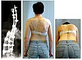
![]() There is no scoliosis without rotation - when the scapulae are at the same height in forward tilt, it is not scoliosis.
There is no scoliosis without rotation - when the scapulae are at the same height in forward tilt, it is not scoliosis.
_(14761624591).jpg)
The rotating bodies of the vertebrae push the ribs on the convex side of the curve dorsally, creating a gibbus. ribs are compressed in the concavity. There are changes in the structures spinee – the bodies of the vertebrae are narrowed to the concave side, the discs are compressed on one side, the pedicles are narrower and shorter, the canal is narrowed (all these changes are mainly typical for idiopathic scoliosis)..
Classification of scoliosis according to etiopathogenesis[edit | edit source]
- Congenital scoliosis
- Idiopathic scoliosis
- infantile
- juvenile
- adolescent
- Neuromuscular scoliosis
- Scoliosis in neurofibromatosis
- Secondary scoliosis
- postural
- in other diseases
- hysterical
Congenital scoliosis[edit | edit source]
The cause is a disorder in the formation or segmentation of the vertebrae embryonically.
- Segmentation disorder – a condition where chorda dorsalis did not divide in a section of varying length:
- in the entire range of the spine - the spine does not grow at all, without curvature,
- only in a certain part - in the place of the non-segmented bar, the spine does not grow, elsewhere it does, which leads to curvature, most often on the thoracic spine.
- Porucha formace – patologický vývoj obratle jako takového – poloobratel, čtvrtobratel, motýlovitý obratel (uprostřed je jakoby zaškrcený).
It can lead to severe deformities or make them mute.
- Mandatory screening – postnatal spine examination.
Therapy[edit | edit source]
Conservative treatment usually fails completely. If the deformity progresses, operative osteotomy osteotomy and spondylodesis are performed at an early age.
Idiopathic scoliosis[edit | edit source]
This is the most common type of scoliosis. With larger curvatures, it deforms the chest and thereby endangers pulmonary and cardiacfunctions.
We divide it according to the time of creation into:
- Infantile type – up to 3 years of life, more in boys, usually corrects spontaneously, more often the curve is left-sided.
- Juvenile type – between 3 and 10 years, both sexes the same, more to the right.
- Adolescent type – between the age of 10 and skeletal maturation, the incidence is the same in both sexes, but heavier curves (above 20°) are mostly in girls.
Therapy[edit | edit source]
Conservative treatment is applied for curves below 40° and for non-progressing curves:
- a curve below 20° – it is monitored and not treated, exercise is recommended,
- curve 20°–40° – plaster corset or Milwaukee corset.
Operative treatment is used for scoliosis with significant progression and for curves above 40°.
- Back procedures - simple compression on the convexity and distraction on concavity, without spondylodesis (artificial fusion of the vertebrae).
- Partial correction using spondylodesis.
- Leading services - removal of discs and fixation of spec. instrumentation.

X-ray image of adolescent idiopathic scoliosis. Thoracic curve 30°, lumbar 53°.

Post-operative X-ray image after correction of scoliosis to less than 15°. Vertebral fusion was performed using wire, screws, hooks, and bone grafts.
Result after scoliosis surgery.


Neuromuscular scoliosis[edit | edit source]
It arises as a result of a developmental disorder with a peripheral motor deficit, when the individual is unable to freely control the muscles from birth. Surgical stabilization and long fusion of the vertebrae are used in the treatment.
Scoliosis in neurofibromatosis[edit | edit source]
A neurofibroma in the spine area causes a short curvature of the spine in the affected section, a compensatory curve is formed in other sections. If possible, the neurofibroma is removed and curve correction with fusion is performed.
Secondary scoliosis[edit | edit source]
They arise due to inflammation (e.g. tuberculosis), injuries, spine operations. Scoliosis can also occur in some diseases – osteogenesis imperfecta, Marfan syndrome, mucopolysaccharidoses, multiple epiphyseal dysplasia.
- Postural scoliosis – when the length of the lower limbs is unequal, a reactive curvature of the spine occurs.
Examination[edit | edit source]

Anamnestically, we determine the development since birth, the general condition of the patient, the family occurrence of malformations and defects, neurological problems, we ask about paini, about menarche.
- Status localis – by aspect, palpation we can find out the direction of the curve, the presence of gibbous,
- to find out the status of hull compensation:
- hang the plumb line from the occiput and when it crosses the gluteal groove, the trunk is compensated,
- the size of the rib prominence can be determined by examination in forward bending,
- we note the height of the shoulders and the slope pans (their asymmetry indicates a decompensation of the curve).
- X-ray examination - standing for a long format to capture the section from the protuberance occ. to to the hip joint, in 2 projections.
Description of scoliotic deformity[edit | edit source]
- The main curve – it is the largest, structural, it arises first.
- Compensatory curve - compensates for the curvature of the spine, goes to the other side than the main one, is above or below the main curve.
- Terminal vertebra – it is placed most cranially or most caudal on the curve, most inclined to the concavity of the curve.
- The apex vertebra is the most rotated vertebra, the most deviated from the vertical curve, the least inclined.
- Curve orientation – means whether it goes left or right.
- Localization - means which part of the spine is affected.
- Weight is measured according to Cobb - it is the angle that the two end vertebrae make together
Links[edit | edit source]
External links[edit | edit source]
- Scoliosis – Diagnosis (CT program)
- Scoliosis of the spine - exercises, exercises, information, types of scoliosis at ZbynekMlcoch.cz
Source[edit | edit source]
- BENEŠ, Jiří. Study Materials [online]. [cit. 2009]. <http://jirben2.chytrak.cz/materialy/orto,trauma_jb.doc>.
References[edit | edit source]
- SOSNA, A – VAVŘÍK, P – KRBEC, M. Fundamentals of Orthopedics. 1. edition. Triton, 2001. 175 pp. ISBN 80-7254-202-8.