Vertebrae
Vertebrae (vertebrae) are the bones that make up the spine. They are connected to each other by fixed and movable spinal connections. The human spine has a total of 33-34 vertebrae.
On each vertebra we distinguish:
- Body (corpus vertebrae) - it faces forward. In the cervical vertebrae, the body is low and gradually increases in size, so that in the lumbar vertebrae the bodies are quite massive.
- The Arch (arcus vertebrae).
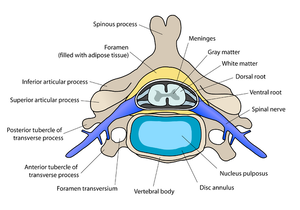
- Processes(processus) – are receding from the vertebral arch. There are a total of seven. The spinous process (processus spinosus) points backwards and is palpable on the back.The two transverse processes (processus transversi) point laterally and have muscles and some ribs attached to them. The two pairs of articular processes point up and down and provide a movable connection between the vertebrae.
The attachment of the vertebral arch to the vertebral body is mediated by a structure called the pedicle (pediculus arcus vertebrae). The arch forms an opening with the vertebral body called the foramen vertebrale through which the spinal cord passes.
Types of vertebrae[edit | edit source]
According to the position of the vertebra on the spine, there are 5 types of vertebrae:
- cervical vertebrae (vertebrae cervicales) – we distinguish 7; C1-C7; C1 (atlas) and C2 (axis) have their specific shape;
- thoracic vertebrae (vertebrae thoracicae) - 12 in total; Th1-Th12 (T1-T12);
- lumbar vertebrae (vertebrae lumbales) - we distinguish 5; L1-L5;
- sacral vertebrae (vertebrae sacrales) - variable number 5-6; S1-S5(6); fused into sacral bone (os sacrum);
- coccygeal vertebrae (vertebrae coccygeae) - variable number 4-5; Co1-Co4(5); Co3-Co4(5) fuse and together with Co1 and Co2 form the coccyx (os coccygis). Co1 and Co2 may be fused cartilage.
Each type of vertebra has a specific shape determined by several structural characteristics.
Cervical vertebrae[edit | edit source]
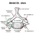
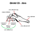
The general structure of the cervical vertebrae is that of C3-C7. The foramen vertebrale is triangular in shape. The facies intervertebralis on the cranial and caudal sides of the vertebrae are elongated ellipsoid in shape, terminating at the lateral ends in a prominence of the uncus corporis vertebrae. Except for C2 (the axis - the condyle), they all contain a closed foramen transversarium through which the arteria vertebralis passes, accompanied by 1-2 venae vertebrales. On this transverse process we identify the tuberculum anterius and t. posterius. The spinous process of the cervical vertebra is forked, characteristically looking like the tail of a swallow. In the middle part there are processus articulares superiores et inferiores, which bear the facies articulares sup. et inf. C7 has a typical prominent spinous process that stands out among the other vertebrae, hence the name C7 - vertebra prominens.
The atlas and the axis (C1 and C2) differ from the general structure of the cervical vertebrae.
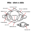
- Atlas lacks the corpus vertebrae, this is replaced by the anterior arch. The arcus anterior and the arcus posterior are then distinguished, which run up to the tuberculum anterius et posterius. On the lateral sides, the massae laterales form the communication between the anterior and posterior arches of C1 and carry cranially the facies articulares superiores et inferiores caudally. On the posterior side, medial to the foramen transversarium lies the sulcus arteriae vertebralis. On the inner side of the anterior arch lies the fovea dentis, which articulates with the dens axis at C2.
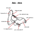
- The Axis, unlike C1, C3-C7, does not form a completely enclosed foramen transversarium. Facies atricularis superiores lie laterally on the body of the condyle and the articular surfaces are separated. On the ventral side of the body, the dens axis projects cranially, replacing the body of the carrier and articulating with the fovea dentis of the carrier.
Thoracic vertebrae[edit | edit source]
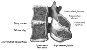
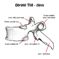
The thoracic vertebrae have a more massive body of semicircular shape, the facies intervertebralis semi-moon-shaped.The processus transversi are dorso-laterally directed and lie directly lateral to the fovea costalis processus transversi for costovertebral articulation. Laterally, at the vertebral body margin, the foveae costales superiores et inferiores, except Th10-Th12, also for the costovertebral articulation. From the processus articulares sup. et inf. protrude the facies art. sup. et inf. for the intervertebral articulation. The medial part of the foramen vertebrale is spherical in shape and projects dorsally, blunt at the end, the processus spinosus.
Lumbar vertebrae[edit | edit source]
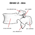
The lumbar vertebrae are characterised above all by their massive kidney-shaped bodies. In the lumbar vertebrae, there are no transverse processes, but processus costales, which are the remains of stunted ribs and point laterally, slightly dorsally. The spinous process of the lumbar vertebrae is massive, blunt and short. Between the spinous and bony processes we find two smaller processes, processus accesories et mammilaries. In this area caudally there is a lower articular process with articular facets and cranially an upper articular process, also with articular intervertebral facets in an almost sagittal plane.
Sacral vertebrae[edit | edit source]
The sacral vertebrae grow into the sacrum (os sacrum). The first sacral vertebra S1, together with the last lumbar vertebra L5, which is inclined caudally in the ventral direction, forms an angular formation called the promontorium.
Coccyx vertebrae[edit | edit source]
The smallest vertebrae in the body are 4-5 in number and can grow together to form the coccyx. The coccyx is connected to the sacrum by a cartilaginous articulation. A fall can dislocate the coccyx. The coccyx plays an important role during childbirth, when extension increases the dimension of the diameter recta apertura pelvis from 9.5 cm to 11.5 cm.
Changes in the number of vertebrae[edit | edit source]
During embryonic development, the number of vertebrae may change, especially in the lumbar region. The change in the number of vertebrae occurs within a range of ±1 by the following phenomena:
- Lumbarisation of the sacral vertebrae (+1 vertebra).
- Sacralization of the lumbar vertebra (−1 vertebra).

