Hip joint
The hip joint, articulatio coxae, is a simple synovial joint that connects the bones of the lower limb and the pelvis. Both hip joints support the trunk and balancing movements contribute to maintaining the balance of the trunk, while allowing movement of the lower limbs relative to the pelvis.
Joint type[edit | edit source]
Joint spherical limited, enarthosis.
Joint surfaces[edit | edit source]
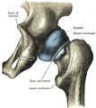
The head is the femur, the caput femoris, which is equal to 3/4 of the surface of the sphere.
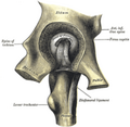
The socket is formed by the acetabulum on the hip bone. It is filled with a fat pad, the pulvinar acetabuli, and extended by a cartilaginous rim, the labrum acetabuli.
Articulating Bushing[edit | edit source]
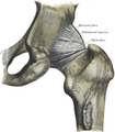
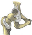
The articular capsule begins at the edges of the acetabulum and attaches to the neck of the femur bone, collum femoris, where it ends anteriorly at the linea intertrochanterica. At the back, it omits the crista intertrochanterica, which serves for the attachment of muscles.
Reinforcement of the joint capsule and joint ligaments[edit | edit source]
The joint capsule is covered by muscles and ligaments, which is why dislocation during an accident is rare.
Iliofemoral ligament
- On the front side of the joint, it runs from the anterior inferior iliac spine to the intertrochanteric line;
- It prevents the trunk from tilting towards the femur, as its tension ends the extension in the joint;
- It is the strongest ligament in the body.
Pubofemoral ligament
- Runs from the pecten ossis pubis to the front of the joint capsule, where it joins the ischiofemoral ligament (see below);
- Limits abduction and external rotation of the joint.
Ischiofemoral ligament
- Located at the back of the joint;
- It starts above the tuber ischiadicum, goes forward over the upper surface of the capsule, joins the ligamentum pubofemorale and together with it forms a fibrous ring in the wall of the capsule that undercuts the caput femoris in the so-called zona orbicularis.
Ligamentum capitis femoris
- A small ligament goes from the lig. transversum acetabuli, closing the incisura acetabuli, to the fovea capitis femoris (the ligament is the remnant of the mesenchyme of the primitive articular disc).
Movements[edit | edit source]
The actual movement of the joint is a rotating movement of the head in the socket, from the basic standing position the following movements are possible:
- flexion – up to 120° (possible increase during simultaneous abduction);
- extension – up to 13° (terminated by tension of the lig. iliofemorale);
- abduction – up to 40°;
- adduction – possible hyperadduction up to 10°;
- rotation – external up to 15°, internal up to 35°.
Flexion, abduction, adduction, and rotation are possible to a greater extent.
Basic and intermediate positions[edit | edit source]
The basic position is when standing upright.
Mid stance is in mid flexion with slight external rotation and abduction.
Blood vessels and nerves[edit | edit source]
Arteries[edit | edit source]
- They arise from the vascular network around the joint;
- one part of the network branches around the acetabulum, receiving branches from a. glutea superior et inferior, a. obturatoria (a. ligamenti capitis femoris), a. pudenda interna and other smaller branches;
- the second part of the network is distinct around the neck of the femur and is supplied by branches from aa. circumflexae femoris, medialis et lateralis , aa. gluteae, superior et inferior and from the deep femoral canal (a. perforans I.);
- from both parts of the net, the branches for the joint case are separated.
Veins[edit | edit source]
- The veins go into the plexus of the capsule and continue along the feeding arteries.
Nerves[edit | edit source]
Innervation:
- front side – n. femoralis;
- medial – n. obturatorius;
- dorsal – n. ischiadicus (plus part of the upper and lateral sides);
- upper and lateral – n. gluteus superior.
Links[edit | edit source]
Related Articles[edit | edit source]
External links[edit | edit source]
References[edit | edit source]
- ČIHÁK, Radomír. Anatomie 1. 2., uprav. a dopl edition. Grada, 2001. 497 pp. ISBN 80-7169-970-5.


