Insulin

| endocrine pancreas | |
| heterodimer composed of two chains (α and β, connected by disulfide bridges) | |
| skeletal muscle, myocardium, adipose tissue, liver | |
| insulin receptor | |
| increases cell glucose entry and proteosynthesis , inhibits glucagon release and MK production; in the liver: glycogenesis , TAG production, glycolysis , decreased glucose and ketone bodies; in muscle: glycogenesis, glycolysis | |
| 176730 |
Insulin is a pancreatic hormone. It is produced in specialized cells of the pancreatic islets - the so-called B-cells. It is a peptide hormone and it regulates energy metabolism. Insulin is also called the hormone of satiety or excess. The diseases associated somehow with insulin (the problem of the synthesis, the problem of islet cells, disorder of receptors and signaling) in our population is relatively common and they are collectively called diabetes mellitus (DM).
Construction of pancreatic islet[edit | edit source]
B-cells (60% of islet cells) are located predominantly in the center, A-cells (25%, produce glucagon), on the contrary, rather on the periphery. The arteriole comes to the center, where it branches and capillaries head towards the edges of the imaginary deformed sphere. In this way, it is possible for the released insulin to act on A-cells, while glucagon usually does not have time to reach B-cells (half-life 1 - 3 minutes, in addition, most of the glucagon is usually taken up by the liver). This is good to note when we talk about regulating hormone secretion.
History[edit | edit source]

As early as 1921, Banting and Best (as the so-called "islet factor") isolated insulin from pancreatic tissue. It belongs to many primacies (among proteins) - it was the first protein where evidence of hormonal activity was given, the first protein that was crystallized and sequenced, the first protein artificially synthesized in the laboratory. On the other hand, we know very little about its intracellular action and various molecular mechanisms.
Structure[edit | edit source]
An insulin molecule is a heterodimer composed of two chains (α and β, connected by disulfide bridges). Throughout the molecule, there are three sites where the amino acid change causes inefficiency - the location of disulfide bonds, the hydrophobic residues of the C-terminus of the β-chain, and the hydrophobic residues of both ends of the α-chain. (Amino acid substitutions are otherwise quite common, but if they occur outside the three sites, they practically do not alter biological efficacy.)
Synthesis[edit | edit source]
Insulin synthesis - like any protein - begins in the nucleus by transcription and continues on ribosomes by RER translation - preproinsulin is formed. It differs from insulin in its pre-sequence (hydrophobic AMK, which serves as a guide for the molecule to travel to the RER tanks) and the linker C-peptide (AMK sequence, which connects the N-terminus of the α-chain and the C-terminus of the β-chain). Pre-sequence is removed in the RER, proinsulin is formed. It has a suitable conformation to oxidize the -SH groups of cysteines to form disulfide bridges between the α- and β-chains. Subsequently, proinsulin is transported to Golgi apparatus, where proteolysis (removal of C-peptide) begins. Insulin, a small amount of proinsulin (insulin / proinsulin ratio is 5: 1), C-peptide and minor amounts of other substances are then packaged into secretory granules and, after an appropriate signal, fuse with the cytoplasmic membrane and release their contents into the extracelullar fluid.
Insulin secretion[edit | edit source]
The secretion takes place through the following mechanism:
- In the case of an increase in the level of glucose in the plasma (state after a meal) - and thus in proportion to the B-cells of the pancreas - there is a change in the membrane potential (depolarization).
- Depolarization is caused by the accumulation of ATP in B-cells, which leads to the closure of ATP-dependent K + channels, so potassium accumulates in the cell. ATP is formed in the respiratory chain, Acetyl-CoA for the citrate cycle comes from the pyruvate dehydrogenase reaction. Pyruvate is formed by aerobic glycolysis.
- Since glucose metabolism begins in B-cells with glucokinase (Km = 10 mmol/l, the same enzyme is still found in hepatocytes), it is necessary for the plasma glucose level to rise to about 8 - 10 mmol/l. This mechanism ensures that large amounts of ATP are formed only at high glucose levels (Glc transporter SLC2A2 is active at higher levels, normal Glc level is 3.6 - 5.5 mmol/l).
- As a result of depolarization, Ca 2+ channels open, calcium levels in the B-cell cytoplasm rise and the cytoskeleton phosphorylates - secretory granules merge with the membrane (Ca 2+ is needed as a cofactor for kinases).
- Postprandial secretion occurs in two "phases" (or two peaks on the plasma insulin level versus time) - the early and late phases.
- It has been observed in mice that insulin secretion is also increased by osteocalcin (a glycoprotein produced by osteoblasts in bone) as well as B-cell proliferation. It is not yet clear in the human body what causes communication between bones and the regulation of energy metabolism. At present, it is clear that the inactivation of the so-called PTPRV gene (encodes a tyrosine phosphatase present in stem cells, Sertoli cells and osteoblasts) has a positive effect on B-cell proliferation and insulin secretion.
- On the other hand, insulin secretion is inhibited by somatostatin.
- In the clinic, it is important to determine the amount of C-peptide as a marker of endogenous insulin production. It is used to distinguish between type 1 and type 2 diabetes, especially in patients treated with exogenously administered insulin.
The mechanism of action[edit | edit source]
The insulin receptor[edit | edit source]
There is a receptor - heterotetramer on the target cell membrane. The α subunit is stored extracellularly, it binds the hormone. The β subunit consists of a transmembrane protein and its intracellular part shows tyrosine kinase activity. The subunits are covalently linked by disulfide bonds in the α 2 -β 2 ratio .
The transfer signal[edit | edit source]
In the case of insulin binding, oligomerization occurs, two (or more) receptors cluster together, and the conformation of the molecules changes, resulting in autophosphorylation of the intracellular portions of the adjacent receptor halves. Receptor synthesis and subsequent degradation occur with a half-life of up to 12 hours. Adapter proteins are used to transfer the signal - in the case of insulin IRS-1 (insulin receptor substrate). The second messenger of insulin is in dispute. The whole cascade ends with phosphorylation / dephosphorylation of target proteins, respectively causes exposure to transport proteins or acts on DNA (see next paragraph). The hormone-receptor complex is also internalized.
Degradation[edit | edit source]
Insulin is degraded (especially in the liver, partly in the kidneys and placenta) by the enzyme insulinase, or glutathione-insulin-transhydrogenase (liver), the receptor is re-exposed on the membrane.
Down-regulation[edit | edit source]
When the insulin concentration is high, the sensitivity of the tissues to insulin decreases (so-called "down-regulation" - reducing the number of receptors on the membranes). This contributes to the development of insulin resistance in DM II.
Insulin action[edit | edit source]

Insulin increases the transport of glucose from the blood to skeletal muscle, myocardial muscle and adipose tissue cells. This is due to the fact that the hormone causes the exposure of the glucose transporters GLUT4 (which have so far been prepared as a whole by the intracellular fluid) on the membrane. This is especially the case in skeletal muscle cells, cardiomyocytes and adipocytes. It has been shown by various methods (subcellular fractionation, electron and fluorescence microscopy) that in the absence of insulin in these tissues, most (approximately 95%) of GLUT4 is located intracellularly.
As insulin is flushed out after a meal - while starving (or several hours after a meal) it drops - glucose is saved for the brain for most of the day (one is generally prepared to experience deficiency rather than excess…) precisely because the muscles and fat does not get, respectively gets a small amount (GLUT4 is missing on the membrane because there is no insulin).
Effects on energy metabolism[edit | edit source]
They are derived from the state in which a person finds himself after eating. The body received a dose of glucose that needed to be processed. Therefore, glycolysis, glycogenesis, lipogenesis and lipid storage in adipocytes will be active . Of course, if necessary, glucose is consumed immediately (for example by working muscle, the brain lives almost only from glucose). Insulin reduces the amount of cAMP, respectively inhibits the conversion of inactive adenylate cyclase to active. Thanks to that:
- protein kinase A is not formed (remains in an inactive state like protein kinase B) that would convert active glycogen synthase to inactive glycogen synthase.
- remains an inactive phosphorylase, which is the control enzyme of glycogenolysis.
- it acts on lipogenesis by activating acetyl-CoA carboxylase and at the same time inhibits lipolysis with low levels of cAMP.
By acting on DNA, insulin induces the biosynthesis of the fatty acid synthase enzyme complex and reduces the synthesis of phosphoenolpyruvate carboxykinase (a regulatory enzyme of gluconeogenesis). In the liver, it inhibits ketogenesis and causes cell growth.
It is good to realize that we have to look at metabolism as such as a big whole. If we affect the metabolic pathway in one cell, it will not go unanswered in other cells. Therefore, on the one hand, we see the direct action of insulin (simply by binding to a receptor in the membrane), and on the other hand, inconspicuous indirect events (for example block lipolysis in adipocytes will cause fatty acids deficiency in the liver, so the liver will process glucose in particular).
In addition, insulin has a positive effect on cell growth and replication, on wound healing (note: in fibroblast cultures, insulin causes the ability of growth factors (FGF, PDGF, EGF…) to stimulate the cell cycle).
Fetal period[edit | edit source]
Insulin begins to form at 10 weeks and also acts on fetal organogenesis. (The placenta is impermeable to insulin, so maternal insulin does not get there).
Insulin in therapy[edit | edit source]

Insulin syringe

Insulin syringe in disassembled form
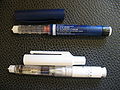
Insulin pen
Insulin pump reservoir


Links[edit | edit source]
Related articles[edit | edit source]
- Insulin therapy
- Hormons of the human body: ADH • Estrogens • Erytropoetin • Gestagens • Glucagon • Glucocorticoids • Human chorionic gonadotropin • Insulin • Catecholamines • Calcitonin • Noradrenaline • Parathormone • Prostaglandins • Renin-angiotensin-aldosteron system • Growth hormone • Testosterone
- C-peptide
- Diabetes mellitus
External links[edit | edit source]
Sources[edit | edit source]
- DUŠKA, František. Biochemie v souvislostech, 1.díl – základy energetického metabolizmu. 1. vydání. Praha : Karolinum, 2006. ISBN 80-246-1116-3.
- MURRAY, Robert K.. Harperova biochemie. 2. vydání. Jinočany : H&H, 1998. ISBN 80-7319-013-3.
- MOORE, Keith L. a PERSAUD. Zrození člověka: embryologie s klinickým zaměřením. 1. vydání. Praha : ISV, 2002. 564 s. ISBN 80-85866-94-3.
- GUYTON, Arthur C. a John E. HALL. Textbook of medical physiology. 11. vydání. Philadelphia : Elsevier Saunders, 2006. 1116 s. ISBN 0-8089-2317-X.
- LENEY, Sophie E. a Jeremy M. TAVARÉ. The molecular basis of insulin-stimulated glucose uptake: signalling, trafficking and potential drug targets. Journal of Endocrinology. 2009, vol. 72, no. 203, s. 1-2, ISSN 1479-6805.