Examination of the chest (pneumology)
Examination of the chest from the perspective of a pulmonologist includes a visual examination , where we mainly notice the abnormal shapes of the chest and the type of breathing. Next, a palpation examination , where we examine chest tremors and pleural friction. By percussion , where we compare the symmetry of the percussion (we detect pathological darkening or hypersonic percussion) and the topographical definition of the lungs. Next, we examine the chest by auscultation , where we evaluate alveolar and tubular breathing - we mainly focus on the intensity of breath murmurs, the ratio of inspiration and expiration and the presence of secondary breath murmurs. By listening, we further investigate the chest voice.
View[edit | edit source]
During the visual examination, we notice the shape, deformities, respiratory movements and soft parts. The shape of the chest changes during growth and puberty. A normal chest is bilaterally symmetrical.
Face[edit | edit source]
We recognize these shapes:
- Barrel-shaped (emphysematous) - found in obstructive lung disease , when expirium is difficult and the chest is in an inspiratory position. All diameters increased (mainly anteroposterior), sternum more arched, spine kyphotic.
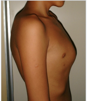
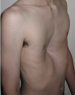
- Pectus carinatum (bird) – chest flattened from the sides, enlarged anteroposterior dimension, sternum pushed forward, e.g. in rickets .
- Pectus infundibuliforme, pectus excavatum (funnel-shaped) – 2 forms: indented lower part of sternum (funnel) or indented entire sternum (boat).
- Kyphoscoliotic – kyphosis and scoliosis of the thoracic spine.
- Deformities - most often the consequences of lung or pleural disease (adhesions after inflammation). Significant deformity can be observed in patients with a congenital heart defect or a defect that arose in early childhood. The bulging heart creates pressure on the chest wall, which creates a hump, also known as a voussûr .
Breathing movements[edit | edit source]
For breathing movements, we monitor the type of breathing, symmetry and breathing frequency (physiologically 16-20 breaths/min). During normal breathing ( eupnoea ), both halves of the chest participate simultaneously and equally abundantly. In men, we observe more of an abdominal type of breathing (mainly movements of the diaphragm), in women, costal breathing (rising and falling of the ribs).
The frequency of breathing can be affected by diseases of the lungs, heart, CNS, anemia or toxic and metabolic influences.
Rapid breathing is called tachypnea . It occurs during agitation, increased body temperature, or increased exertion. Deep breathing is called hyperpnea . Slowed breathing ( bradypnoea ) is found in patients with depression, in patients under the influence of certain drugs, or in patients with increased intracranial pressure. Shortness of breath is referred to as dyspnoea . Temporary cessation of breathing is called apnea .
- Cheyne-Stokes respiration
We find it in heart failure , uremia , severe pneumonia , and increased intracranial pressure (e.g. in CMP ).
- Biot's breathing
In meningitis and encephalitis , when the irritability of the respiratory center is reduced.
- Kussmaul respiration (acidotic)
Tachypnea and hyperpnea are often present. It is most often encountered in diabetic coma (increased amount of ketone bodies ) and metabolic acidosis .
- Sighing
Typical for neurocirculatory asthenia, deepened inhalation with often prolonged expiration. The patient feels that he cannot breathe.
- Orthopnea
Forced position for breathing. It is most often found in patients with lung or heart disease. It is a sitting or semi-sitting position, the hands rest on the mat, the legs are lowered. Venous return to the right atrium is reduced in cardiac patients.
- Breathing with prolonged expiration
Typical for patients suffering from asthma , chronic bronchitis and obstructive pulmonary disease .
Additional attention should be paid to tumors arising from soft tissues, possibly from cartilage and bone . We also pay attention to the examination of the breast , where cancerous growth can occur (mainly in women, but also in men!).
Feel[edit | edit source]
On palpation, we observe resistance, soreness, chest tremors, pleural friction (inflammation of the pleura ).
- Fremitus pectoralis
Or chest tremors. We examine using both palms, which we place on symmetrical places on the chest, which we compare. We invite the patient to say out loud, for example, numbers (1, 2, 3).
- Strengthening is palpable over the infiltrated tissue, such as in pneumonia .
- Weakening or even disappearing occurs with pleural effusion, pneumothorax , adhesions, and blockage of the bronchus by a tumor or foreign body with subsequent atelectasis .
- Pleural friction
Occurs with pleurisy. If it is present, we can feel it during inspiration and expiration. Pleural friction is best palpable at the lower edge of the lungs and in the axillary surfaces.
Percussion[edit | edit source]
In healthy people, the tap is full, clear , comparable on both sides of the chest.
We perform tapping on the back and front of the chest, preferably in a sitting position, or we can film the patient lying down.
We recognize several types of taps:
- Comparison tap
Knocking out the same spots on each side. Sound tap - it's bright, less bright towards the bases.
- Frontal comparison – we knock out and bilaterally compare the area of the supraclavicular pits, parasternal area, medioclavicular area, middle axillary area, usually from top to bottom.
- At the back - we tap paravertebrally from the seventh cervical vertebra (C7), then in the scapular line and in the middle axillary line, again downwards.
- Topographic tap
Delineation of tap change – for example, tap dimming, we proceed perpendicularly to the expected area of tap dimming. Topographical determination of the lower limits of the lungs - in front and on the sides, we orient ourselves according to the ribs and intercostal spaces, in the back according to the vertebral spines.
Physiological limits of the lungs[edit | edit source]
Ventrally - parasternal - 6th rib, in the medioclavicular line - 6th intercostal, in the middle axillary line - 8th rib,
Dorsally – in the scapular line the 10th rib, paravertebrally – the spine of the 10th thoracic vertebra on the right (Th10 P) and the spine of the 11th thoracic vertebra on the left (Th11 L).
Knocking out the diaphragm[edit | edit source]
The diaphragm varies between 4-8 cm depending on the respiratory phase. Bilateral reduction of the displacement of the diaphragm occurs with emphysema , ascites , with an increased condition of the diaphragm during pregnancy , with pleuropulmonary adhesions. Unilateral reduction of the displacement of the diaphragm can be detected in the presence of unilateral thoracic pleural effusion, pneumothorax, pleuropulmonary adhesions, atelectasis of the lower lobe of the lung, palsy of the phrenic nerve , etc.
Tap changes[edit | edit source]
- Hypersonic - in air lungs, in emphysema, in the presence of air - pneumothorax.
- Tympanic - with a large amount of air.
- Darkened to dark - with reduced airiness of the lungs; with thickening of lung tissue – pneumonia, tumor, pulmonary infarction, atelectasis; in pleural thickening; in the presence of fluid in the pleural cavity - fluidothorax (percussion varies according to the amount of effusion).
Listening[edit | edit source]
Listening to the lungs (half heels) - a page with links to recordings of listening phenomena
Under physiological circumstances, breathing above the lungs is cellular, clean, without secondary phenomena . Tubular breathing is audible only above the jugular, upper sternum and between the shoulder blades.
We recognize:
- Direct - placing the ear on the chest.
- Indirect - using a stethoscope.
By listening, we compare both sides, the patient breathes deeply with his mouth open, if possible not loudly.
Basic types of breathing[edit | edit source]
- Cellular respiration
As when exhaling with the mouth set to the letter f , we hear the murmur throughout the inspirium, but in the expirium only at the beginning - ratio inspirium/expirium = 3:1.
- Tube breathing
As in exhaling with the mouth set to the letter ch , physiological breathing for the larynx and trachea region, the expiratory component is greater than the inspiratory component.
Changes in breathing[edit | edit source]
- Enhanced cellular respiration
With increased ventilation - e.g. Kussmaul breathing. It is important when it is unilaterally found in a healthy lung (hyperventilated), when the other lung is affected by, for example, pneumonia or in pneumothorax and inflammatory exudate of the other lung.
- Weakened cellular respiration
Physiologically, it occurs in obese people. Pathologically, we find reduced alveolar respiration in:
- reduction of respiratory excursions, which may occur with chest injury or dry pleurisy ;
- extensive pleuropulmonary adhesions;
- effusion, pneumothorax;
- pulmonary emphysema (reduction of alveoli), when ventilation is reduced;
- in obstructive atelectasis.
- Inaudible breathing
In pneumothorax, increased effusion, or obstructive atelectasis, a large part of the lungs is usually affected.
- Cellular respiration with prolonged expiration
We auscultate in bronchial asthma , in inflammation of the bronchi and bronchioles (resistance in these pathways) – spasm, swelling, secretions and in emphysema (loss of elasticity).
- Pathological tube breathing
Tube breathing where it normally isn't present. For example, when the alveoli are removed from breathing, but the main bronchus is open.
- Elimination of the function of the alveoli:
- filling of cells - inflammatory infiltrate in pneumonia, blood in pulmonary infarction, tumor tissue;
- compression of the cells from the outside - during effusion.
- Compressive breathing (bronchovesicular) - audible above the upper limit of medium and large effusions; is caused by effusion pressure. It is a combination of alveolar and tubular (audible mainly during expiration) breathing.
Secondary breath murmurs[edit | edit source]
They arise during air flow in the presence of viscous or watery secretions in the bronchi, bronchioles or alveoli, or above the pleura under pathological circumstances, also during bronchospasm.
Crops[edit | edit source]
- Dry croup
Viscous, semi-liquid contents (adherent to walls). The vibration of the contents by the air current creates a whistling or creaking sound. It can also be caused by a spasm of the bronchi. It occurs in acute and chronic bronchitis, in bronchial asthma. Wheezes and wheezes are heard in both breathing phases . Wheezes are more likely to be heard in bronchial asthma , wheezing more often in COPD .
- Wet dirt
Liquid to semi-liquid contents. Division into crops of small (cartilage), medium and large bubbles. Sound is produced by the bursting of a bubble on the surface of a liquid. We can divide crops:
- emphatic – clear, coming close; infiltrated, well-conducting tissue – e.g. pneumonia , bronchiectasis ,
- and unstressed - dark, coming from afar; airy, poorly conductive tissue.
Banging[edit | edit source]
Crackling, or crepitus , is only audible during inspiration. Clear, sharp crackles of small bubbles forming a continuous noise, simile − rubbing hair between fingers.
Occurs:
- physiologically – in people breathing shallowly (e.g. after surgery), it disappears after several deep breaths.
- pathologically – with pneumonia, with beginning and ending pneumonia. It can be in pulmonary infarction and infiltrative pulmonary tuberculosis, also in idiopathic pulmonary fibrosis (dry crackles).
Stridor[edit | edit source]
Stridor is a whistling or wheezing phenomenon, the cause is narrowing of the large airways, there is expiratory stridor or inspiratory stridor.
Frictional pleural murmur[edit | edit source]
This sound phenomenon sounds like walking on frozen snow, it is best heard in the axillary region at the base of the lung and at the angle of the scapula, it is characteristic of dry pleurisy, it disappears when an effusion occurs.
Bronchophony[edit | edit source]
Bronchophonia, or chest voice, is investigated by asking the patient to count (1, 2, 3), for example, or to repeat the expression "thirty three". Above a healthy lung, we hear the chest voice very vaguely. The chest voice can be clearly heard over areas of physiological tubular breathing. Above other areas of the lung tissue only when it is pathologically thickened, when its conductivity increases.
- Enhancement – over condensed lung tissue; in pneumonia and pulmonary infarction,
- weakening - it is audible above the effusion, in pneumothorax, atelectasis and in thickening of the pleura.
Links[edit | edit source]
References[edit | edit source]
- KLENER, Pavel, et al. Propedeutics in internal medicine. 2nd edition. Prague: Galén, Karolinum, 2006. 320 pp. Chapter 10.3.: Examination of the chest. pp. 85-92. ISBN 80-7262-429-6 .
- CHROBÁK, Ladislav, et al. Propedeutics of internal medicine. 2nd edition. Prague: Grada, 2007. 243 pp. Chapter 5.: Examination of the chest. pp. 51-66. ISBN 978-80-247-1309-0 .
Reference[edit | edit source]
- CHROBÁK, Ladislav, et al. Propedeutics of internal medicine. 2nd edition. Prague: GRADA Publishing, 2007. 243 pp. ISBN 978-80-247-1309-0 .


