Ankylosing spondylarthritis

| Risk factors | HLA-B27, male |
|---|---|
| Classifications and references | |
| MKN-10 | M45, juvenile ankylosing spondylarthritis M08.1 |
| MeSH ID | D013167 |
| OMIM | 106300 |
| MedlinePlus | 000420 |
| Medscape | 332945 |
Ankylosing spondylarthritis (also morbus Bechtěrev or spondylitis ankylosans) is a spondylarthritis, a group of inflammatory rheumatic diseases affecting connection on the spine (intervertebral, costovertebral, SI joint, discs and ligaments of the spine, sometimes root or peripheral joints). It leads to the gradual ossification of the joint capsule and ligaments and thus to the ankylosis of segments up to the entire spine (it can stiffen in any position).
Etiopathogenesis[edit | edit source]
The disease begins at a young age (usually the 2nd-3rd decade). It is more common in men. The disease is linked to HLA-B27. The etiological agent has not been demonstrated. The role of intestinal microflora is considered.
Symptoms[edit | edit source]
The symptoms of the disease are divided into three groups: axial, peripheral and extra-articular symptoms.
Axial symptoms[edit | edit source]
The basic symptoms of the disease include inflammatory pain in the lower back - it arises on the basis of sacroiliitis, typically in the second half of the night and wakes the patient from sleep. It is accompanied by morning stiffness. Both pain and stiffness improve with warming up. The pain spreads to the higher areas of the spine (except for the cervical spine) - spondylitis is a treasure. Axial symptoms also include arthritis of the shoulder and hip joints.
Peripheral symptoms[edit | edit source]
Asymmetric oligoarthritis most often develops with a preference on the lower limbs. Dactylitis (so-called sausage finger ) can also occur - affecting the interphalangeal joints of one finger and tendon.
Extra-articular symptoms[edit | edit source]
- eyes - acute anterior uveitis,
- heart - aortic insufficiency, transmission disorders, aortitis,
- GIT – ulcerative colitis, Crohn's disease,
- lungs - fibrosis,
- kidneys – amyloidosis,
- vertebral osteoporosis
Diagnostics[edit | edit source]
Physical examination[edit | edit source]
During the examination, we focus on the sacroiliac joints, the development of the spine in three planes and the expansion of the chest.
- Mennel's maneuver - We push the patient's hip bones. The test is positive if the patient feels pain on the injured side.
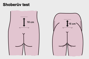
- Schober test - Shows the development of the lumbar spine.
- Forestier flèche - This is the distance of the occiput to the perpendicular wall. It should be a maximum of 2 cm.
- lateral flexion
- chest expansion - at least 5 cm
Imaging methods[edit | edit source]
X-rays are the first manifestation of sacroiliitis. Furthermore, syndesmophytes and ossification of tendon attachments arise, which leads to the fusion of vertebral bodies to the image of the so-called bamboo stick . Sacroileitis is often bilateral.
We distinguish 5 stages according to the location of changes.
| Stadium | inflammatory changes |
|---|---|
| Stadium I | unilateral sakroiliitida |
| Stadium II | bilateral sacroiliitis |
| Stadium III | lumbar spine involvement |
| stadium IV | thoracic spine involvement |
| stadium V | cervical spine involvement |
Another important imaging method is magnetic resonance imaging, where a typical finding is the presence of effusion or swelling of the bone marrow.
Laboratory tests[edit | edit source]
In the laboratory finding we find an increase in sedimentation and CRP, normocytic normochromic anemia.
Treatment[edit | edit source]
The basis of treatment is regular, lifelong exercise, rehabilitation and physical therapy. Spa treatment is indicated for patients every year. Pharmacological treatment includes:
- nonsteroidal antirheumatic drugs,
- DMARDs – effective only sulfasalazine in forms with peripheral arteritis,
Links[edit | edit source]
Related articles[edit | edit source]
- Juvenile idiopathic arthritis
- Rheumatoid arthritis
- Psoriatic arthritis
- Manifestations of inflammatory rheumatic diseases on the musculoskeletal system and their surgical treatment
References[edit | edit source]
- PASTOR, Jan. Langenbeck's medical web page [online]. ©2010. [cit. 02-06-2010]. <https://langenbeck.webs.com/>.
- ČEŠKA, Richard, et al. Interna. 1. edition. Praha. 2010. 855 pp. ISBN 978-80-7387-423-0.
References[edit | edit source]
- ↑ ČEŠKA, Richard, et al. Interna. 1. edition. Praha : Triton, 2010. 855 pp. ISBN 978-80-7387-423-0.


