Transport across cell membrane
Cell membrane is semipermeable. Substances that pass through it can pass freely or with the help of membrane carriers. Transport of the substances can then be active or passive.
Passive Transport[edit | edit source]
Passive transport is the transfer of substances across the cellular membrane, which takes place spontaneously through channels and proteins. Unlike active transport, this event does not consume any chemical energy (ATP). Passive transport depends on the permeability of the cell membrane, which is composed of a double layer of phospholipids and interspersed proteins. The basic types of passive transport are simple diffusion, facilitated diffusion and osmosis.
Diffusion[edit | edit source]
Diffusion is a spontaneous process of penetration of particles of one substance into another with an effort to uniform penetration into the entire volume. It occurs because of the disordered thermal movement of particles. Substances tend to move from an environment of higher concentration to an environment of lower concentration. Diffusion is not an energetically demanding event. Diffusion enables the movement of substances inside cells and thereby metabolism.
Simple Diffusion[edit | edit source]
Simple diffusion enables the transport of substances along the concentration gradient (from places with higher concentration to places with lower concentration). It takes place with polar molecules of small dimensions or various types of gases.
Facilitated Diffusion[edit | edit source]
Facilitated diffusion is a type of passive transport in which substances cross the membrane along their electrochemical gradient using carriers built into the membrane.
Osmosis[edit | edit source]
Osmosis is a type of passive transport in which a solvent (most often water) moves through a semipermeable membrane from a space with a less concentrated solution to a space with a more concentrated solution.
Permeation through ion channels[edit | edit source]
Ion channels along with transporter proteins are structures that participate in transports across the biological membrane. We can divide them according to the principle of their opening.
- Ion channels still open
- Voltage-gated ion channels
- Chemically gated ion channels
- Voltage and Chemically Controlled Ion Channels
- Mechanically controlled ion channels
Active transport[edit | edit source]
"Active transport" is the transfer of substances across the cellular membrane, which, unlike passive transport, is linked to energy consumption. Thanks to the supplied energy, which is most often produced by the splitting of ATP, it is possible to carry out this transport also against the direction of the concentration gradient (concentration gradient).
Active transport is enabled by specialized integral membrane proteins embedded in the cell membrane:
- "Ion pumps" – ion channels equipped with ATPase enzyme.
- Transport proteins equipped with an ATPase enzyme.
Ion pumps[edit | edit source]
Ion pumps are penetrating integral proteins in the cell membrane that serve to transport substances against the concentration gradient. During the transfer of substances there is an ATP consumption.
Endocytosis[edit | edit source]
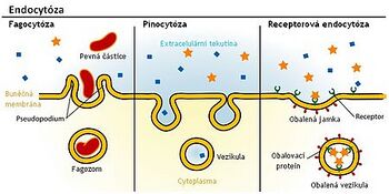
Endocytosis is an energy- and material-intensive process characteristic of animal cells. During endocytosis, there is absorption of particles from the external environment. Cells are separated from the external environment by a cytoplasmic membrane. Some hormones, lipoprotein particles, viruses, antibodies, but also damaged cells or bacteria get into them through endocytosis.
Phagocytosis[edit | edit source]
Phagocytosis is the ability of cells to absorb foreign particles, microbes or damaged cells.[1] Cells capable of phagocytosis participate in the nonspecific immunity of the organism - antigen presenting cells, monocytes, from which individual types of [[Macrophages|macrophages] develop ] (Kupffer Cells, histiocytess, microglia and others), and white blood cells (neutrophil leukocytes, eosinophilic leukocytes).
Pinocytosis[edit | edit source]
Pinocytosis is one subtype of endocytosis. During pinocytosis, the cell receives ``extracellular fluid (extracellular fluids = ECF) and very small particles.
Exocytosis[edit | edit source]
Exocytosis is a continuous process in which the cell "excretes" through the cell membrane (plasmalemma) larger particles (eg macromolecules) directly "into the extracellular matrix". The membrane vesicle (vesicle) containing the secretion travels to the membrane, fuses with it and subsequently releases the internal contents into its surroundings.
Links[edit | edit source]
Related Articles[edit | edit source]
References[edit | edit source]
- ↑ ŠVÍGLEROVÁ, Jitka. Phagocytosis [online]. [cit. 2011-01-02]. <http://wiki.lfp-studium.cz/index.php?title=Fagocyt%C3%B3za&oldid=137>.
Source[edit | edit source]
- ŠVÍGLEROVÁ, Jitka. Internet archive [online]. [cit. 2010-11-12]. <https://web.archive.org/web/20160306065550/http://wiki.lfp-studium.cz/index.php/Passivní_transport>.
- ŠVÍGLEROVÁ, Jitka. Internet archive [online]. [cit. 2010-11-12]. <https://web.archive.org/web/20160306065550/http://wiki.lfp-studium.cz/index.php/Facilitovaná_difuze>.
- ŠVÍGLEROVÁ, Jitka. Internet archive [online]. [cit. 2010-11-12]. <https://web.archive.org/web/20160306065550/http://wiki.lfp-studium.cz/index.php/Osmóza>.
- CODEC, M. – KARPENKO, V.. Biophysical Chemistry. 1st edition. Prague : Academia, 2000. ISBN 80-200-0791-1.
- VAJNER, Luděk – CARBON, George – KONRÁDOVÁ, Wenceslas. Medical histology I. 1st edition. Prague : Karolinum Publishing House, 2012. ISBN 978-80-246-1860-9.
- LEOŠ NAVRÁTIL, ROSINA JOZEF AND COLLECTIVE, Medical biophysics [online]. [feeling. 2014-16-11]. <[https://www.grada.cz/medicinska-biofyzika-
- LANGMEIER, Miloš. Fundamentals of Medical Physiology. 1st edition. Grada Publishing, a.s, 2009. pp. 320. ISBN 978-80-247-2526-0.
- KONRÁDOVÁ, Václava. Functional Histology. 2nd edition. H + H, 2000. pp. 291. ISBN 978-80-86022-80-2.
- HALL, J.E. Textbook of Medical Physiology. 12th edition. Philadelphia : Saunders Elsevier, 2011. ISBN 978-1-4160-4574-8.
- BALOUNOVÁ, Z. Plant Physiology [online]. [cit. 2010-11-16]. <http://kbd2.zf.jcu.cz/text/lidi/balounova/fros/FYZR0712.ppt>.
- ALBERTS, B. Molecular Biology of the Cell [online] . 4th edition. New York : Garland Science, 2002. Available from <https://www.ncbi.nlm.nih.gov/books/NBK26896/>. ISBN 0-8153-3218-1.
- MESCHER, Anthony L. Junqueira's basic histology: text and atlas. Thirteenth edition. New York [etc.]: McGraw-Hill Medical, 2013. ISBN 978-0-07-178033-9.
- WORSE, Wenceslas – BARTŮŇKOVÁ, Dahlia. Fundamentals of immunology. 3rd edition. Triton, 2008. pp. 280. ISBN 80-7254-686-4.
- BROOKER, Robert. Biology. 2nd edition. New York : McGraw-Hill Science/Engineering/Math, 2011. ISBN 9780073532240.
- ALBERTS, Bruce – BRAY, Dennis – JOHNSON, Alexander. Fundamentals of Cell Biology. 2nd edition. Ústí nad Labem : Espero Publishing, 1998. ISBN 80-902906-2-0.
- TRKANJEC, Z. – DEMARIN, V .. ScienceDirect [online]. [cit. 2014-11-26]. <http://www.sciencedirect.com/science/article/pii/S030698770091260X>.


