Ion channels
Ion channels, together with carrier proteins are structures involved in transport across biological membranes.
Structure[edit | edit source]
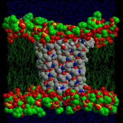
The channel is composed of five protein subunits that pass through a double layer of phospholipids. In the case of the acetylcholine-controlled (cholinergic receptor) channel, it opens when acetylcholine binds to the binding site. Very few channels open without the action of acetylcholine. The ion channel is highly selective, due to the negatively charged amino acid chains. Therefore, the channel only releases positively charged ions such as K+, Na+.
The principle of opening channels[edit | edit source]
A channel in a closed conformation has its passage closed by a so-called gate, which does not let anything through. Binding of acetylcholine causes the subunits, including the gate, to be delayed. At this point, Na+ or K+ can pass through the membrane along the gradient of its electrochemical potential.
Non-gated channels[edit | edit source]
Always open ion channels are water-filled labyrinths rapidly changing configuration and electrical charge. Ions move through them according to the concentration gradient and membrane potential (they have a charge). This type of ion channel has highly selective permeability for one or more ions or molecules. The selectivity depends on the characteristics of the channel and its internal surface.
Voltage controlled ion channels[edit | edit source]
Voltage-gated ion channels open and close with a change in the electrical potential across the membrane. This is due to a configurational change in the protein that makes up the channel.
- A strong negative charge on the inside of the cell membrane leads to the closure of the channel.
- When the negative charge begins to decrease on the inside of the membrane the ion channel opens.
These ion channels work with a certain delay. This is characteristic of them.
Chemically controlled ion channels[edit | edit source]
The change in permeability here is due to the reaction between the receptor and the ion channel.
- The receptor is an immediate part of the channel.
- Activation of the receptor induces G-protein-coupled reactions that lead to phosphorylation of the channel.
- Activation of the receptor induces G-protein-coupled reactions that alter the cellular concentration of metabolic factors but do not lead to phosphorylation of the channel.
- Receptor activation is directly transferred to the ion channel via the G-protein.
G-proteins are GTP-binding regulatory proteins that mediate transmission from a variety of receptors to ejector molecules (e.g. ion channels). They are heterotrimers composed of α,β,γ subunits. The α subunit has the ability to bind GTP or GDP and has intrinsic GTPase activity. Mainly this subunit carries specific properties of G-protein types and interacts with both the receptor molecule and the effector molecule.
This type of channel control occurs, for example, in neurons in the synaptic cleft.
Voltage and chemically controlled ion channels[edit | edit source]
Voltage and chemically controlled ion channels open upon membrane depolarization. The probability and duration of opening depends on the receptor interactions.
Mechanically controlled ion channels[edit | edit source]
Mechanically controlled ion channels are sensitive to cytoskeletal tension. They are part of a large series of mechanoreceptors. Stretching the cell membrane leads to the opening of the ion channel. This mode of regulation occurs, for example, in the organ of Corti in the cochlea of the inner ear, where sound vibrations vibrate the basilar membrane, causing the stereocilia to tilt. Stereocilia (apical specialization of cells) are protrusions of hair cells. Each stereocilia is connected by a filament to another stereocilia. Tilting the filament causes an ion channel in the membrane to open, causing K+ to enter, which in turn stimulates the auditory nerve. This mechanism is very sensitive; the softest sound will move the fibres by 0.04 nm.
Links[edit | edit source]
Related articles[edit | edit source]
Used literature[edit | edit source]
- LANGMEIER, Miloš. Základy lékařské fyziologie. 1. edition. Grada Publishing, a.s, 2009. 320 pp. ISBN 978-80-247-2526-0.

