Shockwave
Template:Checked A shock wave is a physical phenomenon, during which excitement (energy) spreads through the environment in the form of a sudden change in physical quantities describing the state of the environment. field (e.g. electromagnetic).
Pressure shock wave arises during explosive events: during an electric discharge in a liquid, an explosion, a supernova explosion, etc. The source of a pressure shock wave is also objects moving faster than the speed of propagation of the excitation in the given environment, e.g. during supersonic speed of movement of the aircraft or the tip of the whip. In this case, it is a sonic boom.
The passage of a strong enough shock wave can change the properties of the environment itself - in that case the shock wave is referred to as "strong". E.g. in the case of a pressure shock wave, the state or structure of the material through which the wave passes may change as a result of the passage of a "strong shock wave". The shock wave can also cause chemical reactions in the medium.
Pressure Shock Wave[edit | edit source]
Most of the shock waves that the reader can normally encounter are waves of compression and rarefaction of the environment, i.e. pressure shock waves.
These shock waves are created, for example, in the air during a lightning discharge (thunder), during an explosion or a shot. However, the sound itself is not a shock wave, as it usually has a fairly long duration and a de facto constant speed in the given environment. However, the shock wave itself is characterized by a sudden and discrete change in the properties of the medium (environment) in which it propagates and loses its energy when passing through the given environment. For the purposes of this text, the term "shock wave" will refer to the shock wave of pressure.
Difference between shock wave and ultrasound[edit | edit source]
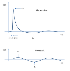
Densification and rarefaction of the environment is the mechanism by which shock and ultrasonic waves propagate in the environment. In the case of ultrasound, however, it is a periodic event with lower amplitudes and a continuous course of pressure. For clarity, you can remember what ripples look like on a pond after throwing a stone.
If we try to transmit large amplitudes of pressure (necessary to crush stones) into the tissues using ultrasound, the negative pressure phase of the ultrasound wave will cause the formation of cavitation and the growth of microbubbles. Gases are present in low concentrations in body fluids and tissues in the natural environment. The thrust phase of the ultrasound wave causes a negative pressure in the liquid (tissue). In the resulting negative pressure, the microbubbles expand and the dissolved gas from the surroundings begins to diffuse into them. The subsequent pressure phase of the ultrasound wave compresses these microbubbles again. With intense ultrasound, we de facto pump energy into a cloud of oscillating bubbles. These oscillating bubbles form an obstacle in the propagation of intense ultrasound to greater depths, but they are used with success, e.g. in ultrasonic cleaners and disintegrators (e.g. for homogenization of suspensions).
Shock Wave, on the other hand, is aperiodic and non-linear. It propagates through the environment as one strong pressure pulse, with a non-linear, almost "jump" pressure increase at the wave front, usually followed by a longer and lower vacuum oscillation. Shock waves generated by a spark discharge in water have a P+ pressure amplitude of the order of 100 MPa (100x more than atmospheric pressure), a P- vacuum amplitude of about 5-10 MPa, and a rise time of 30-120 ns. [1], [2]
If we were again looking for an analogy with waves on water, then the shock wave can be compared to a tsunami wave in a certain approximation. The negative pressure phase of the shock wave has a far (~10x) smaller amplitude (and longer duration) than its pressure phase. This is why the shock wave is more suitable for transferring large pressure amplitudes to the stone. However, even the negative pressure phase of the shock wave can cause the formation of cavitation and microbubbles. However, the time between two shocks is incomparably longer than the time between the following ultrasound wave. The microbubbles therefore have time to relax and shrink more before the next impact due to the diffusion of gases through the bubble wall. The remaining microbubbles no longer form a substantial ex. for the propagation of the following shock wavethe whole thing.
In conclusion, let's summarize the essential features of RV:
- shock waves for clinical stone crushing are focused;
- RV have a significantly larger (>10x) amplitude of P+ pressure than P- negative pressure;
- RV are applied repeatedly, but with a delay sufficient to relax the environment.
Shockwave Generators[edit | edit source]
Three types of shock wave generators have been developed for non-invasive lithotripsy – electrohydraulic, electrodynamic and piezoelectric generators.
- Electrohydraulic - A pressure wave is caused by the formation of plasma by the action of an electric current in water.
- Piezoelectric - They are based on indirect piezoelectric effect = deformation of certain materials due to electric voltage.
- Electrodynamic - The principle is the conversion of the electrodynamic pressure of the magnetic field into acoustic pressure when the electric impulse passes through the coil.
- Effect of Focused and Radial Extracorporeal Shock Wave Therapy on Equine Bone Microdamage
- Effectiveness of radial shockwave therapy compared to focused shockwave therapy
Medicinal use of RV[edit | edit source]
Lithotripsy - breaking up body concretions[edit | edit source]
Extracorporeal shock wave lithotripsy (LERV) is a non-invasive method used to remove (break up) kidney or gallstones. The goal of the therapy is the destruction/fragmentation of stones in the body with minimal damage to the surrounding soft tissue. The shock wave is generated outside the patient's body by a shock wave generator (see Shock wave generators) and passes with minimal losses through the water environment and soft tissues to the stone. The energy of the shock wave is released on the surface of the stone, creating tensile forces on the surface of the stone caused by the reflected energy. Energy that would pass through the stone acts on its far side. At the same time, cavitation caused by negative pressure forces acts on the surface of the stone. The stone is broken into small fragments by the action of the mentioned forces and is naturally washed out of the body in the case of kidney stones, or further dissolved by drugs in the case of gallstones.
Shock waves are generated in water, which has an acoustic impedance comparable to the acoustic impedance of soft tissues, and there is no significant energy dissipation at the interface of the two environments (on the surface of the patient's body). However, if the stone is resistant and a lot of shock waves are needed, petechial bleeding and subcutaneous hematoma can rarely occur on the surface of the body.
Focusing of shock waves contributes to the non-traumatic transfer of high energy to the concretion inside the patient's body. Shock waves are generated outside the patient's body and are focused by reflection or refraction so that the maximum pressure (focus of the shock wave) is in the place where we need to destroy the stone. Thus, waves with a relatively low intensity pass through the tissues, and the destructive effect of the wave is concentrated only at the focal point.
Use of RV in the treatment of musculoskeletal diseases[edit | edit source]
While in non-invasive lithorypse we try to ensure that the RV only passes through soft tissues, this is not the case in musculoskeletal treatments.
Treatment of sprains and slow-healing fractures[edit | edit source]
The first attempts to use shock waves in orthopedics were focused on the treatment of back jointss. Patients with slow-union fractures and with arthrodesis can be successfully treated with high-energy shock waves (such as those used in noninvasive lithotripsy). After exposure of the fusion surfaces with a shock wave, the fracture is immobilized. In more than 80% of cases, the first therapy already results in bone consolidation and at the same time a reduction of symptoms. Side effects are local reactions (swelling, hematomas, petechial bleeding). The treatment is non-invasive and technically easy to perform. In 2001, the authors of a study of 100 patients concluded that the use of extracorporeal shock wave therapy should be the method of first choice in the treatment of patients with clubfoot and slow-healing bone fractures. [3]
Other orthopedic indications for RV treatment[edit | edit source]
- Calcifying tendinitis - Painful shoulder syndrome
It is a disease of the tendons of the shoulder joint of unclear etiology, which is characterized by the deposition of calcium salts in the rotator cuff. Shockwave therapy improved mobility, reduced pain, and reduced the size of calcifications in a randomized trial. The effects of high-energy shock waves were demonstrably better than low-energy RV effects, but they were also more effective than placebo. The effect can be considered proven, even if the mechanism of action remains unclear. [4]
- Epicondylitis of the lateral humerus - Tennis elbow
Epicondylitis is a disease of the tendon attachments (enthesopathy) of the elbow. The cause is usually overuse of the elbow joint during sports or at work. Without treatment, difficulties can persist for a very long time (months or even years).
- http://josr-online.biomedcentral.com/track/pdf/10.1186/1749-799X-7-11?site=josr-online.biomedcentral.com
- https://josr-online.biomedcentral.com/articles/10.1186/1749-799X-7-11,
- http://asadl.org/jasa/resource/1/jasman/v120/i5/p3064_s3?bypassSSO=1
Cardio
- http://www.amscl.com/main.files/paper/2010%20Double-Blind%20and%20Placebo-Controlled%20Study%20of%20the%20Effectiveness%20and%20Safety%20of%20Extracorporeal%20Cardiac%20Shock%20Wave%20Therapy%20for%20Severe%20Angina%20Pectoris.pdf
- http://www.sciencedirect.com/science/article/pii/S0167527309015198
Course of therapy[edit | edit source]
The whole procedure takes around 15 minutes and consists of 3 stages:
- Locating painful places by touch.
- Applying the gel.
- Shockwave application.
Usually, the therapy is repeated 3-5 times, with an interval of 3-10 days.
RV Application Methods[edit | edit source]
- Mapping - used in the initial phase of therapy and serves to:
- acclimatization of the patient to the sensation caused by acoustic waves;
- achieving the greatest possible analgesic effect in the most painful area;
- supporting blood circulation in the given area.
- Rotation - used in the main phase of therapy and serves to:
- focus on the most painful point;
- achieving the maximum therapeutic effect at the key point of the patient's illness.
Clinical Use[edit | edit source]
- Rehabilitation - shoulder pain, epicondylitis, low back pain, Achilles tendon pain, patellar tendinitis, trigger point treatment
- Orthopedics - frozen shoulder, periarthritis humeroscapularis, calcar calcanei/ heel spur, arthritic changes - secondary symptoms, achillodynia, lateral and ulnar epicondylitis
- Sports medicine - muscle distension, prolonged treatment of joint distortion, groin pain, hip pain, low back pain, achillodynia
- Aesthetic medicine - cellulite removal, skin regeneration
Contraindications to RV treatment[edit | edit source]
- Current treatment with anticoagulants (anticoagulant treatment). These are preparations of the coumarin type (e.g. Warfarin, Lawarin, etc.) or current treatment with preparations of the heparin type (LMWH - e.g. preparations Clexane, Fraxiparine, Zibor, etc.).
- Congenital and acquired bleeding and coagulation disorders or bleeding manifestations. In these cases, shock wave treatment may induce some local bleeding manifestations.
- Pregnancy, bacterial infection of the bone (infected fractures, osteomyelitis) or patients with unhealed wounds, Alzheimer's disease, polyneuropathy, in patients with malignant tumors and in patients in whom the exact cause is not determined their musculoskeletal pain or the pain is not precisely localized.
- After the injection of cortisoneoids in the previous treatment (e.g. application of preparations Diprophos, DepoMedrol, etc.), shock wave treatment is suitable only 6 weeks after the last application.
Reimbursement by insurance companies[edit | edit source]
The price of one application (one use) of shock waves is around CZK 400-600 depending on the affected area and is NOT covered by public health insurance
References[edit | edit source]
- ↑ McClure S, VanSickle D, White R: Extracorporeal shock wave therapy: What is it? What does it do to equine bone. Am Assoc Equine Practnr 46: 197–199, 2000, online:http://kiropraktik.dk/GraysonFinalReport.pdf
- ↑ MCCLURE, Scott and Christian DORFMULLER. Extracorporeal shock wave therapy: Theory and equipment. Clinical Techniques in Equine Practice. 2003, year 2, No. 4, pp. 348-357. ISSN 15347516. DOI: 10.1053/j.ctep.2004.04.008. Available from: http://linkinghub.elsevier.com/retrieve/pii/S1534751604000137
- ↑ Schaden, W., A. Fischer, and A. Sailler, Extracorporeal Shock Wave Therapy of Nonunion or Delayed Osseous Union. Clinical Orthopedics and Related Research, 2001. 387: p. 90-94.
- ↑ Gerdesmeyer L, Wagenpfeil S, Haake M, et al. Extracorporeal Shock Wave Therapy for the Treatment of Chronic Calcifying Tendonitis of the Rotator Cuff: A Randomized Controlled Trial. PIT. 2003;290(19):2573-2580. doi:10.1001/jama.290.19.2573.


