Membrane transport
Membrane transport is the transfer of substances through:
1. biological membrane[edit | edit source]
- selectively passes molecules that can pass through the phospholipid bilayer via protein transporters or across the membrane continuum
- the transfer of substances takes place between the cell and the extracellular space or between organelles
- we distinguish:
2. nuclear envelope[edit | edit source]
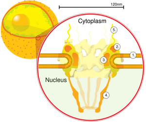
- it takes place through holes, the nuclear pores, which have a diameter of 70 nm, are lined with 8 proteins called nucleoporins, from which fibrils of the nuclear pore, which have the function of receptors, extend on the cytoplasmic side. On the inside is the nuclear basket, which selectively passes molecules into the karyoplasm.
- this is how RNA and protein macromolecules pass through the nuclear pores
Pasive transport[edit | edit source]
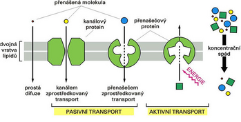
It allows the penetration of substances through the cell membrane without energy consumption in the direction of the concentration gradient from places with a higher concentration to places with a lower concentration. The speed of penetration is influenced by the size of the gradient, temperature, size of the surface - the cell increases its surface area of projections (microvilli, stereocilia...) and folds (invagination)
1. diffusion
- simple – penetration of substances directly through the lipid bilayer (mainly non-polar molecules for which the phospholipid membrane is not an obstacle and small gas molecules, e.g. O2, CO2)
- facilitated – through transport proteins that are characterized by their selectivity and ability to close (channels controlled by voltage, ligand, mechanically or randomly).
2. osmosis – penetration of water molecules through the plasma membrane depending on the environment in which the cell is located (hypertonic, isotonic, hypotonic).
- water does not pass through the lipid bilayer directly, but with the help of special protein carriers - aquaporins
Active transport[edit | edit source]
Energy-intensive transport of substances using ATP via:
1. membrane continuum
- pinocytosis – intake of substances in the form of a solution (cell drinking). Tiny protrusions of the cell's cytoplasm surround a small amount of extracellular fluid and form
pinocytic vesicles. Actin microfilaments are involved in the formation of pinocytic vesicles. These vesicles can merge with early endosomes and lysosomal transport vesicles (the contents of pinocytic vesicles are further processed in this way) or are used for transcytosis.
- phagocytosis – intake of larger solid particles (cell eating). The particle is captured on the surface of the cell membrane by cytoplasmic protrusions. This creates a phagocytic vacuole, which further merges with early endosomes and lysosomal transport vesicles. A cell can absorb foreign material - heterophagy or the organism's own damaged cells or their parts - autophagy. Some self cells (mainly T lymphocytes) are able to cross the membrane continuum of another cell without damaging each other - this is referred to as peripolesis or emperipolesis. The cytoplasmic membrane of cells of the immune system does not contain only its own products. Cells come into contact and often part of the membrane of the neighboring cell is carried away. This is called trogocytosis.
- exocytosis – the opposite of endocytosis, excretion of harmful and waste substances, release of proteins that are part of the extracellular space or substances affecting various functions in the body, e.g. hormones
2. ion channels equipped with ATPase – ion pumps – create a concentration gradient.
Active transport by ion pumps[edit | edit source]
1. Primary active transport[edit | edit source]
It serves to transfer substances against their gradient, consumes energy from ATP or other high-energy phosphate bonds (creatine phosphate CP - in muscles; derivatives of pyrimidine and purine bases - guanosine triphosphate GTP, cytidine triphosphate CTP, ...).
The substances transferred in this way include sodium, potassium, calcium, hydrogen and other ions. For example, restoration of the resting potential after nerve excitation is ensured by Na+-K+-ATPase.
Primary active transport uses different types of ATPases:
- P-type ATPase: sodium-potassium pump, calcium pump, proton pump
- F-ATPase: mitochondrial ATP synthase, chloroplast ATP synthase
- V-ATPase: vacuolar ATPase
- ABC (ATP binding cassette) transporter: MDR, CFTR, …
2. Secondary active transport[edit | edit source]
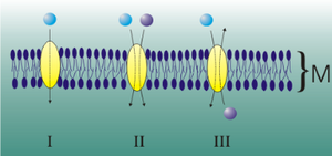
- It connects the movement of several molecules
- cotransport – transports two or more molecules in the same direction = symport;
- opposite (counter) transport – transports molecules in the opposite direction = antiport.
Another possibility is that the gradient created by the transfer of one molecule allows the transfer of another molecule against its gradient. An example is glucose transport in kidney tubules. Sodium cations are pumped by the Na+-K+-ATPase from the inside of the cell extracellularly into the interstitium (energy consumption). This results in a relative increase in their concentration in the urine compared to the cell cytosol. As a result, sodium cations can penetrate back into the cell through a spontaneous exergonic process (during which there is a conformational change ensuring easier binding of glucose).
Types of pumps[edit | edit source]
Sodium-potassium pump[edit | edit source]
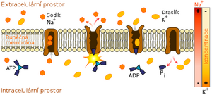
(also Na+ /K+ -ATPase) is an integral membrane enzyme from the class of hydrolases ensuring the counter-directional primary active transport of Na+, K+ ions. At the expense of hydrolysis, 1 ATP molecule transports 3 Na+ ions from the cell and 2 K+ ions into the cell. It is present in all plasma membranes. It plays an important physiological role especially in kidney cells and nerve cells, where it restores the resting potential (-70 mV) after the passage of the action potential (+40 mV) during the refractory phase of the neuron (the time during which the cell is unexcitable).
- It draws sodium from the intracellular space to the extracellular one.
- It draws potassium from the extracellular space into the intracellular space.
Structure[edit | edit source]
The pump consists of two subunits – alpha and beta. Both subunits are proteinaceous substances that pass through the cell membrane. The alpha subunit transports ions and has ATPase activity. On the intracellular side there are binding sites for Na+ and ATP, on the extracellular side there are binding sites for K+. The beta subunit probably anchors the pump in the cell membrane.Alpha and Beta subunit of sodium-potassium pump
Transport mechanism[edit | edit source]
Inside the cell, 3 sodium cations and one ATP molecule bind to the binding sites of the alpha subunit, the cleavage of which enables a change in the conformation of the pump. Sodium is released extracellularly. The new shape has a high affinity for potassium ions. 2 bound potassium cations and their subsequent release intracellularly cause restoration of the original conformation. In nerve cells, up to 70 % of their energy can be consumed by this pump.
Calcium pump[edit | edit source]
In a normal situation, calcium ions outside the cells are in about 10,000 times higher concentration, this level inside the cell is ensured by calcium pumps in two places:
- on the cell membrane – transports calcium cations out of the cell;
- on the membranes of cell organelles (mainly sarcoplasmic reticulum) in muscle tissue – transports Ca2+ cations back to the sarcoplasmic reticulum - the pump is referred to as SERCA 1 in skeletal muscles (Sarcoplasmatic or Endoplasmatic reticulum Ca2+ ATP-ase 1). SR is then an important source of Ca2+ for the initiation of muscle contraction.
- It works on the same principle as the Na-K pump, it has a receptor for Ca2+ and an active ATPase site.
Hydrogen/Proton Pump[edit | edit source]
Occurs:
- On the inner membrane of mitochondria;
- maintains a proton gradient between the intercrystalline and intracrystalline spaces;
- during the transition of H+ back into the matrix, the energy is used to create ATP (respiratory chain).
- In early endosomes - the action of proton pumps lowers the pH, which is used in receptor-mediated endocytosis when, after the fusion of the vesicle with the early endosome, the bond between the receptor and the ligand is released at a low pH (around 5). Receptors are concentrated in the part of the vesicle that is first removed and transported back to the cytoplasmic membrane. The remainder of the vesicle is called the late endosome;
- In the gastric glands:
- Here the pumps are the most active in the whole body, thanks to them HCl is secreted into the stomach in such a way that at the secretory end of the parietal cells in the gastric glands, the concentration of H+ is increased about a million times as a result of the work of these pumps, and then these ions are released into the stomach together with chloride ions anions – formation of HCl.
- In the distal tubules and cortical collecting ducts of the kidney:
- Excess hydrogen cations are transported from the blood into the lumen of the canals (into the urine) - thereby also maintaining the acid-base balance of the organism (acidifying the urine).
- Excess hydrogen cations are transported from the blood into the lumen of the canals (into the urine) - thereby also maintaining the acid-base balance of the organism (acidifying the urine).
Related articles[edit | edit source]
Links[edit | edit source]
Used sources[edit | edit source]
- KODÍČEK, Milan – KARPENKO, Vladimír. Biofyzikálna chémia. 1. edition. Vydavatelství VŠCHT, 1997. ISBN 80-7880-273-1.
- KONRÁDOVÁ, Václava, et al. Funkční histologie. 2. edition. H + H, 2000. 291 pp. ISBN 978-80-86022-80-2.
- HALL, J.E – GUYTON, A.C. Textbook of Medical Physiology. 12. edition. Philadelphia : Saunders Elsevier, 2011. ISBN 978-1-4160-4574-8.
- BALOUNOVÁ, Z. Fyziologie rostlin [online]. [cit. 2010-11-16]. <http://kbd2.zf.jcu.cz/text/lidi/balounova/fros/FYZR0712.ppt>.
- ALBERTS, B, et al. Molecular Biology of the Cell [online] . 4. edition. New York : Garland Science, 2002. Available from <https://www.ncbi.nlm.nih.gov/books/NBK26896/>. ISBN 0-8153-3218-1.
- VAJNER, Luděk, Jiří UHLÍK a Václava KONRÁDOVÁ. Lékařská histologie. 1, Cytologie a obecná histologie. 1. vydání. Praha : Karolinum, 2010. 110 s. ISBN 978-80-246-1860-9


