Lymphadenopathy


Lymphadenopathy is a state of which the dominant feature is local or generalized enlargement of lymph nodes accompanied by other symptoms. It is a frequent working diagnosis, which requires following differential diagnosis with the aim to find the underlying etiology.
Diagnosis[edit | edit source]
The first them is anamnesis with the emphasis on the speed of growth of the lymph nodes, pain, injury and other affects in the drained area of the node. Concomitant symptoms include fevers and especially night sweats and weight loss. Next we inquire about contacts with animals, being abroad (especially developing countries with low hygienic standard, sexual contacts. Pharmacological anamnesis is important aswell.[1]
In the course of physical examination, during palpation of lymph nodes, we are interested in approximal size (millimeters), consistency (fluctuations, toughness), moveability in relation to the base, palpative painfulness. Next we investigate if there is a solitary node or a packet or generalized lymphadenopathy. We also notice if the finding is symmetrical. During aspection we notice the state of the surrounding skin and mucosae. We bear in mind the inspection of all lymph node areas.
From the basic laboratory tests we indicate FW, blood count with differential count, biochemistry - sodium, pottasium, chlorides, creatinine, urea, bilirubin, liver function tests, glucose, CRP, orientational urine tests.[1]
Valuable information of exact dimensions, vascularization and relations to the surroinding structures can be attained by ultrasonography.[2] Uzliny v supraklavikulární oblasti nejsou za fyziologického stavu hmatné vůbec.[2]
If we do not reveal the cause by the means mentioned above, the node (in the case of a packet many nodes) can be surgically extirpated and examined.
![]()
Differential diagnosis[edit | edit source]
Basic causes of lymphadenopathy can be subdivided into inflammatory, caused by a tumor and others.
- Inflammatory
- Infectious
- Viral: EBV, CMV, HSV, adenovirus, enterovirus, rubella a measles, HIV, HHV-8;[3]
- bacterial: pyogenous streptococci, staphylococcus aureus.[3]
- Noninfectious
- Tumors
- Primary include malignant lymphoproliferations;[1]
- Secondary include metastases of solitary tumors.[1]
Sarcoidosis
- Sarcoidosis, thesaurismoses, systemic diseases of connective tissues (SLE, RA, Sjögren's syndrome), hyperthyreosis.[1]
Gallery[edit | edit source]

Neck lymphadenopathy in a patient with EB virosis

RTG of pulmonary hilar lymphadenopathy
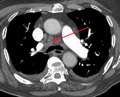
Mediastinal lymphadenopathy (CT)


Links[edit | edit source]
Relevant articles[edit | edit source]
References[edit | edit source]
- ↑ a b c d e Incomplete citation of article. NAVRÁTIL, Milan. Uzlinový syndrom, praktické poznámky k diferenciání diagnostice a diagnostickému postupu. Interní medicína ve zkratce [online]. 2003, no. 5, p. 27-29, Available from <https://www.internimedicina.cz/pdfs/int/2003/01/08.pdf>. ISSN 1803-5256.
- ↑ a b BALLOVÁ, Veronika. Uzlinový syndróm [online]. [cit. 2017-10-21]. <http://www.vpl.sk/files/file/XXXI_conf_w/onkologia/Uzlinovy%20syndrom.pdf>.
- ↑ a b CHOVANEC, Martin – KOMÍNEK, Pavel – ZELENÍK, Karol. Příručka pro praxi : Diferenciální diagnostika krčního uzlinového syndromu [online]. ©2014. [cit. 2017-10-21]. <http://www.otorinolaryngologie.cz/dokumenty/PPP_lymfadenopatie.pdf>.