Periodontal trunk
From WikiLectures
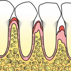
A periodontal trunk (pocket) as a manifestation of periodontal disease can only occur in one tooth. It is usually found in multiples as a result of chronic periodontitis, which is the most common cause of stumps.
It is characterized by the deepening of the gingival sulcus due to the loss of attachment in the region of the neck and root of the tooth. As a result, the slit-like space between the gum and the tooth widens.
The probeable proboscis depth is physiological up to 3.5 mm. If the trunk is deeper, it is pathological.
- The root surface in the region of the periodontal trunk is covered with bacteria, endotoxins and altered infected cementum.
- The gingival wall of the trunk is covered with non-keratinizing squamous cell epithelium, the subepithelial layer is infiltrated by leukocytes.
- The bottom of the trunk is formed by a narrowed layer of connective epithelium (compared to a healthy one).
- The contents of the trunk are subgingival calculus, subgingival plaque, detached epithelia, microbes, leukocytes, pus, granulation tissue.
Division[edit | edit source]
- True periodontal trunk - it is caused by loss of tooth attachment, it is deeper than 3.5 mm.
- False periodontal trunk - there is no loss of attachment (the relationship of the bottom of the trunk to the cementoenamel border is physiological), the depth of the trunk is determined by the extension of the gingiva in the coronal direction. In the case of secondary infection of the periodontal trunk, a right trunk may occur.
- Hypertrophy of the gingiva occurs, for example, after some drugs (Cyclosporin A, hydatoinates, antiepileptics, dihydropyridine blockers of Ca2+ channels - antihypertensives).
According to trunk depth[edit | edit source]
- Supraalveolar - the bottom of the trunk is located above the edge of the alveolar bone. Most often in horizontal bone resorption.
- Infraalveolar - the bottom of the trunk is below the edge of the alveolar bone. Vertical bone resorption.
- Deep trunks are deeper than 5.5 mm.
Diagnostics[edit | edit source]
- Survey.
- X-ray examination: OPG, intraoral.
Therapy[edit | edit source]
- periodontitis therapy;
- modification of medication (usually not possible) - surgical removal.
Links[edit | edit source]
Related Articles[edit | edit source]
Source[edit | edit source]
- MÁZANEK, George – URBAN, Francis, et al. Stomatological refresher course. 1. edition. Prague : Grada Publishing a.s, 2003. 456 pp. ISBN 80-7169-824-5.
- LOGENÍK, Paul. Professional practice of a dentist : comprehensive guide to dentistry. Part 6. 1. edition. Prague : Dashöfer, publishing house, 2004. ISBN 80-86229-21-1.

