Laboratory indicators of iron deficiency or excess
Serum iron determination[edit | edit source]
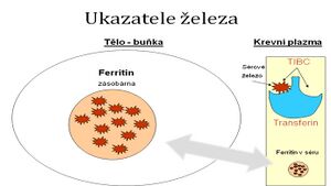
The FE transit pool is determined as plasma or serum iron. Transferrin binds virtually all iron in the circulation.
Normal ferremia:
- newborns: 17.90–44.75 μmol/l,
- children: 8.95–21.48 μmol/l,
- women: 7.16–26.85 μmol/l,
- men: 8.95–28.64 μmol/l.
Reduced values: with iron deficiency – intestinal resorption disorder, reduced food intake, increased losses (most often through menstrual bleeding , but also latent losses to the digestive tract from various causes), increased need (pregnancy, hyperthyroidism).
Increased values: in hemolytic anemias and hemochromatosis.
Plasma transferrin[edit | edit source]
Physiological concentration: 2–3 g/l
- An iron transport protein that is formed in the liver and is subsequently released into the circulation.
- Increase - with a lack of iron in the body, the feedback mechanism increases transferrin synthesis in hepatocytes .
- Decrease - in case of hepatic proteosynthesis disorder, malnutrition, losses ( proteinuria, exudative enteropathy), acute or chronic inflammation (negative acute phase reactant ).
Transferrin saturation[edit | edit source]
Transferrin saturation is sometimes given as % iron saturation.
Calculation: serum iron: TIBC ( total iron binding capacity )
Norma: 20–55 %.
Reduced values: with iron deficiency.
A decrease in saturation along with low TIBC values accompanies hemosiderosis, hemochromatosis, liver disease.
Increased values: 80–90% in hereditary hemochromatosis.
Serum ferritin[edit | edit source]
We use this value to assess the status of iron reserves in the body. It has no function in terms of Fe metabolism.
Norm: (μg/liter; ng/ml)
- newborn: 25–200,
- 1st month: 200-600,
- 6 months–15 years: 7–140,
- women: 12–150,
- men: 15–200.
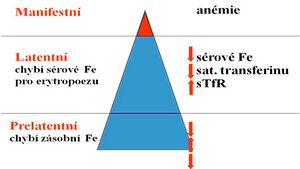
A decrease in the level of serum ferritin is already demonstrable in the initial stages of iron deficiency anemia.
Increase: with iron overload, liver damage , tumors; is an acute phase protein.
Serum (soluble) transferrin receptor (sTfR)[edit | edit source]
Determined by immunoanalytical methods, most often by ELISA
Increase: iron deficiency in the body, increased expression of the transferrin receptor on cell membranes, e.g. during intensive hematopoiesis (hemolytic anemia, β-thalassemia , polycythemia ).
Reduction: bone marrow suppression , chronic renal failure .
Note: The transferrin receptor (TfR) is the main mediator of iron uptake into the cell. TfR is a transmembrane dimer of two identical subunits that binds and then internalizes transferrin with two iron molecules. Thus, the transport of iron into the cell cytosol is enabled. As the need for iron in the cell increases, the expression of this receptor on the cell membrane increases (approximately 80% of all TfRs are on erythroid progenitor cells). Soluble transferrin receptor is formed by proteolysis of TfR at a specific site of the extracellular domain. This results in a monomer detectable in plasma or serum. There is a constant relationship between TfR and sTfR, therefore sTfR is an indirect indicator of TfR expression in the organism.
Erythrocyte ferritin[edit | edit source]
- Its amount reflects the status of iron reserves during the last 3 months (Fe deficit/overload).
- Not affected by acute illness or liver function.
Free erythrocyte porphyrin[edit | edit source]
Increased in disorders of heme synthesis .
| Anemia of chronic diseases | Iron deficiency anemia | Myelodysplastic syndrome | |
|---|---|---|---|
| Serum Fe | ↓↓↓ | ↓↓↓ | ↑↑↑ |
| Transferrin/TIBC | ↓ | ↑↑↑ | ↓↓↓ |
| Ferritin | ↑ | ↓↓↓ | ↑↑↑ |
| Bone marrow | Fe 2+ v MF | Missing Fe | Round sideroblasts – Fe reserves ↑ |
| Dr. clues | The underlying disease | Symptoms ↓ Fe bleeding | Dyshaematopoiesis |
Links[edit | edit source]
Related articles[edit | edit source]
References[edit | edit source]
NEČAS, Emanuel. Obecná patologická fyziologie. 2. edition. Prague : Karolinum press, 2007. ISBN 978-80-246-1291-1.


