Inflammatory rheumatic diseases
Rheumatic diseases are among the most widespread human diseases. They occur in up to 30% of the population, for 3% they mean permanent disability.
Rheumatoid arthritis
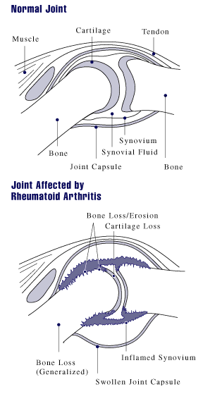
Rheumatoid arthritis (polyarthritis progressiva primaria chronica) is a chronic inflammation characterized by synovial hypertrophy with infiltration by inflammatory cells, destruction of articular cartilage and decalcification of bone (osteoporosis). Rheumatoid arthritis is characterized by the production of antibodies (RF – rheumatoid factor, ANF – antinuclear factors) and acute phase proteins. Clinically, rheumatoid arthritis can be decribed as symmetrical polyarthritis, which predilectionally affects the small joints of the hand and radiocarpal joints, with prolonged morning stiffness.
Occurence
Rheumatoid arthritis is 2-3 times more common in women. Symptoms develop most often between the ages of 20 and 40. However, there is also juvenile rheumatoid arthritis, which primarily affects large joints.
Etiology
Rheumatoid arthritis is an autoimmune inflammation that is often associated with the HLA-DR4 or DR1 immunophenotype. The initiator is believed to be an as yet unknown microbial pathogen (EBV, retroviruses, parvoviruses, Borrelia etc. are being considered). It is not known exactly which antigen the autoimmune reaction is directed against – probably against collagen type II or against cartilage glycoprotein 39 (it binds to DR4). Rheumatoid factor occurs in 80% of patients, i.e. antibodies against the Fc fragment of IgG.
Pathological-anatomical changes
The first changes are in the synovium, then in the fluid, in the cartilage and finally in the para-articular. First, serofibrinous intra-articular inflammation develops, the pannus forms. Pannus is a villous overgrown synovial membrane, in which ligaments and blood vessels proliferate excessively. It covers the articular cartilage, seperating it from the nourishing synovial fluid. As a result, chondrocytes disappear and subchondral bone mass is gradually eroded. If the pannus growing from opposite sides of the joint joins, it can further fibrotic change, ossify, and finally ankylosis occurs.
Another morphological manifestation of rheumatoid arthritis are rheumatic nodules, which arise mainly in the subcutaneous tissue . Histologically, they consist of three layers:
- centrally there is necrotic tissue,
- around it is a layer of palisade fibroblasts and multinucleated cells,
- peripherally there is a layer of chronic inflammation.
Clinical picture
Joint disorders
- Symmetric polyarthritis
Initially, the joints of the hand are affected (starting from the periphery → proximal interphalangeal → „bottle fingers“; metacarpophalangeal; radiocarpal). It usually does not affect the distal interphalangeal. Later, ulnar deviation of the fingers, swan neck deformities (hyperextension in the proximal interphalangeal joint and flexion in the distal interphalangeal joint) and buttonhole deformities (flexion in the proximal interphalangeal joint and hyperextension in the distal interphalangeal joint) are typical. The joints are painful at rest, during palpation and movement, there is morning stiffness (it takes more than an hour to move). The joints have the classic signs of inflammation in addition to redness. In more severe cases, chronic inflammation can lead to volar subluxation of the wrist and rupture of finger tendons. The activity of the process is fluctuating, often depending on the weather.
- Damage to individual joints
- Elbow disorders - flexion contractures.
- Shoulder joints rotator cuff ruptures.
- Hip joints - affected less often.
- Knee joints – angular deformities and flexion contractures. Fluid may enter the popliteal bursa – Baker's cyst. On the leg, hammertoes and hallux valgus are typical findings.
- Spine
Affected mainly in the neck region, the involvement of the atlantoaxial joint with subluxation is severe (neck and head pain, spinal cord compression). Sudden death can also be a complication of subluxation.
![]()
- Impairment of the temporomandibular joint
It causes pain when chewing.
Course of the disease
There are 3 types of the course of the disease:
- monocyclic – one cycle of the disease followed by remission lasting more than 1 year;
- polycyclic – slowly progressing course with episodes of incomplete remissions (most common);
- progressive – permanent progression without remissions.
Extra-articular disability
The disease can be accompanied by extra-articular disabilities:
- rheumatoid nodes (under the skin, especially above the elbows and above the proximal edge of the ulna), usually multiple, usually painful nodules, up to several cm in size;
- tendosynovitis (mainly in the hands, tendon ruptures with the development of deformities - swan neck, buttonhole);
- osteoporosis (initially periarticular, later diffuse - pathological fractures);
- secondary amyloidosis (AA, especially kidney damage);
- hematological abnormalities (mainly anemia, thrombocytosis);
- eye disorders (iritis, iridocyclitis, keratoconjunctivitis);
- damage to the skin, heart, blood vessels, nerves, lungs, etc.
Diagnostics
Laboratory finding
- Inflammatory markers (↑ FW, CRP).
- Antibodies:
- rheumatoid factor (RF) – antibody (mostly IgM) against Fc fragments of IgG, demonstrated by latex-fixation test;
- anti-CCP – antibody against cyclic citrullinated peptide, are more specific for RA than rheumatoid factors;
- APF – antiperinuclear factors;
- ANF – antinuclear factors.
- Punctate (biochemically RF, high content of polymorphonuclear cells).
X-ray changes
- Early – swelling of soft tissues near the joints, periarticular osteoporosis, marginal bone erosion.
- Late – narrowing of the joint space, diffuse osteoporosis, deformities, bone ankylosis.
Four stages according to Steinbrocker were introduced for the evaluation of X-ray images.
| Stage | Characteristics |
|---|---|
| Stage I | periarticular osteoporosis, no destruction |
| Stage II | slight signs of destruction, no deformities |
| Stage III | destruction of cartilage and bone, deformities |
| Stage IV | fibrous or bony ankylosis |
- Furthermore, scintigraphy can be used in the diagnosis (it will show the distribution of the disability in individual joints).
Criteria for diagnosis
For the diagnosis of rheumatoid arthritis, the presence of 4 of the 7 criteria is important:
- morning stiffness;
- arthritis of 3 or more areas;
- arhtritis of the joints of the hand (RC, MCP, PIP);
- symmetrical arthritis;
- rheumatoid nodules;
- rheumatoid factor (RF);
- X-ray changes.
Therapy
Regime measures
In the acute stage, bed rest, prevention of contractures, antalgic splints, etc.
Physical therapy and rehabilitation
Maintaining the range of motion in the joint, preventing muscle weakness.
Pharmacotherapy
The basis of pharmacological treatment is disease modifying antirheumatic drugs (DMARDs = disease modifying antirheumatic drugs).
Disease-modifying drugs (DMARDs)
This includes two groups of drugs:
- conventional,
- biological treatment.
- Conventional drugs
- metothrexate – the most frequently used drug, the drug of first choice,
- leflunomid – inhibitor of pyrimidine nucleotides, has effects similar to methotrexate,
- sulfazaine,
- hydroxychlorine and chlorochine – have the weakest effect.
- Biological treatment
- TNFα inhibitors – etanercept, infliximab, adalimumab, golimumab, certolizumab pegol
- rituximab – a chimeric monoclonal antibody against the CD20 molecule,
- abatacept – blocks T-lymphocyte activation by blocking the costimulatory signal,
- tocilizumab – an antibody against the receptor for IL-6,
- anakinra – IL-1 receptor antagonist.
Other medications
- non-steroidal antirheumatic drugs and analgetics – only symptomatic drugs – COX1 inhibitors – diclofenac, indometacine, selective COX2 inhibitors – nimesulid, coxib;
- corticoids – generally (prednisone) or intra-articularly (triamcinolone) – to bridge the period until the onset of effect of DMARDs.
Surgical treatment
- synovectomy (possibly also by radiation application of yttrium isotope to the joint);
- total endoprosthesis;
- arthrodesis (fixation of the joint in a favorable position, pain relief, most often the radiocarpal area).
Summary video
Bechterev's disease

| Risk factors | HLA-B27, male |
|---|---|
| Classifications and references | |
| MKN-10 | M45, juvenile ankylosing spondylarthritis M08.1 |
| MeSH ID | D013167 |
| OMIM | 106300 |
| MedlinePlus | 000420 |
| Medscape | 332945 |
Children's Rheumatoid Diseases
Rheumatology surgery
- The operation often ends in failure, the cooperation of an orthopedist, a rheumatologist and a rehabilitation worker is necessary;
- the primary task is the prevention of deformities - synovectomy;
- other tasks:
- to correct created deformities (arthroplasties, arthrodesis, alloplasty),
- restore lost joint functions, relieve pain;
- wound healing (vasculitis) is often poor;
- poor access to intubation in spinal disorders is often negative for surgery;
- worse course of infections;
- a long-term operational plan is being developed - operating on individual joints (in stages).
Links
References
- ČESKA, Richard, et al. Internal 1st edition. Prague: Triton, 2010. 855 pp. ISBN 978-80-7387-423-0 .
References
- BENEŠ,. Study Materials [online]. [cit. 2011]. <http://jirben2.chytrak.cz/>.


