Histochemistry
This is a histological method in which we demonstrate the presence of substances (e.g. enzymatic activity) in the sample using a chemical reaction. It deals with the morphology of cells, but also describes the chemical substances in cells and demonstrates cell inclusions. In histochemistry, we demonstrate the presence of e.g. polysaccharides, lipids, enzymes.
Conventional histochemistry[edit | edit source]
Evidence of inorganic ions and compounds[edit | edit source]
Demonstration of certain substances in the body is diagnostically important, whether in the case of forensic medicine (here, for example, due to poisoning - As, Pb, Hg, Ag) or in pathology due to deviations from the norms for the occurrence of substances (Ca, Fe, Zn, Al).
- Ca occurs in the body in soluble, insoluble, ionized and non-ionized form. It is proven to be ionized, for example, due to HE staining blue in an alkaline reaction (pH > 9).
- Fe3+ is demonstrated using the Perls reaction (see below).
- Zn as a component of insulin or as a cofactor of many enzymes is demonstrated by zincone with a blue result, dithizone with a red result
Perls reaction[edit | edit source]
Siderophages (macrophages) engulf old or damaged erythrocytes. Degradation of heme produces iron ions in them (we can also find them in ferritin), which are stored in the storage form of iron - hemosiderin. After the addition of yellow blood salt (potassium hexoxycyanoferrate) and HCl (to create an acidic environment), the reaction with hemosiderin produces a blue precipitate known as Berlin blue (~ Prussian blue).
The Perls reaction is therefore used to detect hemosiderin, which is physiologically found in large quantities only in siderophases. It also serves to distinguish lipofuscin, hematoidin, and hematin, which are Perls negative.
- Methodology of staining
The composition of the coloring solution
- potassium ferrocyanide
- Distilled water
- hydrochloric acid (2%)
The sections are placed for 30 minutes in the staining solution at a temperature of 60 °C. The sections are then removed from the staining solution and counterstained with nuclear red or hematoxylin.
PAS reaction[edit | edit source]
The PAS (P'eriodic Acid Schiff) reaction is based on the oxidation of free hydroxyl bonds, e.g. the 1,2-glycol bond between two adjacent carbons in hexoses , using periodic acid (HIO4). Aldehyde groups are formed, which react with Schiff's reagent (basic fuchsin + sodium pyrosulfite Na2S2O5) to form a new complex compound that it is purple in color.
Structures that can be detected by this method are called PAS positive (e.g. glycogen in the liver).
- Methodology of staining
Schiff's reagent composition:
- basic fuchsin (=pararosaniline)
- Distilled water
- sodium pyrosulfite
- concentrated hydrochloric acid
The tissue section is oxidized in 1% periodic acid for 10 minutes. They are then removed, rinsed in water and stained with Schiff's reagent for 10 minutes. Finally, the section is rinsed in distilled water and stained with hematoxylin (10 minutes).
Best's crimson is used as a method of detecting glycogen in a place with too high a concentration, where the PAS method is not clear.
Feulgen Reaction[edit | edit source]
This is a reaction that demonstrates the presence of DNA. It consists in the hydrolysis of DNA with hydrochloric acid, whereby the purine bases are cleaved from the carbohydrates and thus the aldehyde groups on the deoxyribose are exposed. Then, similar to polysaccharide detection, the aldehyde groups react with Schiff's reagent to form an insoluble purple precipitate.
The method is used, for example, in pathology in tumor diagnostics to determine cell polyploidy.
Proof of lipids[edit | edit source]
Lipids are demonstrated on frozen sections because the dye has a greater affinity for fat in the tissue than for the substance in which it is dissolved. Dyes are therefore dissolved in organic solvents (isopropanolol, propylene glycol, etc.), which must be sufficiently diluted. Dyes used to represent fats include Sudan III and IV (red) and Sudan black (black), as well as oil red or Nile blue (distinction between acidic and neutral lipids). Fats must not be washed out of the tissues during processing. We use Baker's fluid (water, formalin, calcium chloride) as a fixative – it reduces the solubility of non-polar lipids.
Evidence of phospholipids - we stain with Luxol blue. A method suitable for the representation of the myelin sheath of nerve fibers
Catalytic histochemistry[edit | edit source]
This method is quite demanding, while it uses the basic principle that the enzyme reacts with the substrate, which is converted into the final product of the histochemical reaction. Only this product is then converted into a colored compound (visualization reaction). This principle is used in many applications, i.e. from antibody markers or hybridization probes (see below), through the detection of metabolic processes in the cell, to pathology and forensic medicine . During all these procedures, 4 basic rules for performing catalytic histochemistry must be observed:
- Accuracy - while maintaining the morphology of the monitored cells or tissues, the product must not diffuse and must remain in places where the enzyme is expected to occur.
- Specificity - the final product is the result of the reaction of only one expected enzyme. This is verified on control sections.
- Reproducibility - the experiment can be repeated without significant deviations.
- Validity - the enzyme must not be lost when manipulating the tissue, its distribution and activity are preserved.
To preserve the function of the enzyme, the tissue cannot usually be fixed
Affinity Histochemistry[edit | edit source]
One of the younger fields, which is increasingly used both in research and diagnostics. It allows the detection of very small amounts of substances in the target tissue.
Immunohistochemistry[edit | edit source]
It uses the basic principle of the antigen–antibody interaction, where antibodies specifically bind to the target antigen against which they were "raised". Monoclonal antibodies are produced by hybridomas. Polyclonal antibodies arise after immunization in the organism of a person or animal. Both types of antibodies are used in immunohistochemistry. At the same time, they tend to be labeled for some reason, and this results in the visualization of the site of interaction and the detection of antigens.
Either direct labeling of the antibody is used or the unlabeled antibody is visualized using another labeled antibody:
- Direct reaction - in this case, the primary antibodies that are labeled attach to the antigen. It marks the site of interaction, but the disadvantage is low sensitivity and the need to use an increased amount of antibodies.
- Indirect reaction, ABC reaction, PAP reaction, etc.- avoids the disadvantages of direct reaction and increases the sensitivity of the reaction. The primary antibody attaches to its antigen and the secondary antibodies bind to the primary antibodies. The antigen is marked with a much higher number of marker molecules, which increases the sensitivity of the reaction.
Markers used to visualize the reaction include:
- fluorochromes – to make the reaction visible in a fluorescence microscope
- avidin-biotin complex - a complex often used for signal amplification (ABC method)
- autoradiography – antibodies carry radioactive substances that emit radiation onto the photographic emulsion
- enzymes – catalytic histochemistry is used; the antibody carrying the enzyme attaches itself to the antigen and after delivery of the substrate, the enzyme converts it into a colored product, e.g. PAP (peroxidase-antiperoxidase) reaction
The use of immunohistochemistry enables, for example, the visibility of cytoskeleton filaments, hormones, receptors, etc.
Lectin histochemistry[edit | edit source]
Lectins are proteins (or glycoproteins) that are used, for example, to determine blood groups, mitogenic stimulation of lymphocytes or normal cells. It binds highly specifically to the carbohydrate part of macromolecules.
In situ hybridization[edit | edit source]
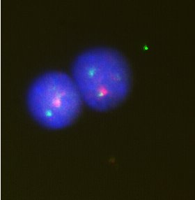
If a certain sequence of nucleotides in DNA or mRNA is known, it is possible to prepare a marked piece of sequence (so-called probe/probe) complementary to the original sequence of nucleotides. Based on base pairing, the labeled probe then attaches to the complementary section of DNA/mRNA and makes it visible. It is used for and to identify chromosomes in interphase, very often as FISH (fluorescence in situ hybridization).
Links[edit | edit source]
Related articles
- Principles of conventional histochemistry in light microscopy
- Staining in light microscopy
- Hematoxylin-eosin staining
References
- MAŇÁKOVÁ, Eve – SEICHERTOVÁ, Alexandra. Methods in Histology. 1. edition. Prague : Karolinum, 2002. 54 pp. ISBN 80-246-0230-X.
- JUNQUIERA, L.Carlos – CARNEIRO, Jose – KELLEY, Robert O. Fundamentals of Histology. 1. edition. Jinocany : H & H, 1997. 502 pp. pp. 14-25. ISBN 80-85787-37-7.


