Endotracheal intubation


Endotracheal intubation is the process of inserting a tracheal tube into the trachea. It allows for realiable securing the airway to ensure patency and suction of secretions.
Indication for intubation[edit | edit source]
Indications for intubation partially overlap with other methods of securing the airway. Compared to less invasive methods, it mainly has the advantage of ensuring adequate ventilation and a lower risk of aspiration. Long-term endotracheal intubation is usually replaced by tracheostomy.
- Respiratory protection against aspiration (unconsciousness including GA, intoxication, stroke, disorders of neuromuscular transmission, ...).
- Obstruction of airways (trauma, bleeding, foreign body, infection, abscess, hematoma, edema, anaphylaxis, laryngospasm, secretions).
- Shortness of breath with necessity artificial lung ventilation (acute respiratory distress syndrome, pneumonia, complicated asthma, COPD, pulmonary edema, ...)
- Disorders of lung mechanics (chest trauma, pneumothorax, fluidothorax, ...)
- Shock states.
- Specific cases with the need for UPV (hyperventilation in intracranial hypertension, preventive intubation in patients at risk of deterioration for transport, ...)
- Among the indications for securing the airways for general anesthesia for operations, it is primarily the following conditions where we prefer intubation over, for example, laryngeal mask:
- The patient is not hungry (shock, pregnancy, ascites, reflux, pyloric stenosis).
- Laparoscopic procedures using capnoperitoneum (intra-abdominal pressure increases the risk of aspiration).
- Performances on the head or neck.
- Abdominal or chest procedures.
- Surgery with the patient in the prone position.
- For operations with a planned duration of more than 45 minutes.
Contraindications are associated with the necessity of surgical securing of the airways tracheostomy or coniotomy - this is primarily a complete obstruction of the upper airways and loss of facial injuries.
Execution[edit | edit source]
Division by approach[edit | edit source]
- Orotracheal (always with urgent intubations, more frequent),
- Nasotracheal (newborns, small children, better fixation of the tube, dental surgery).
Division by display method[edit | edit source]
Direct laryngoscopy we equalize the anatomical conditions and allow a direct view into the larynx using a laryngoscope. Laryngoscopes are divided according to the shape of the spoon into bent (standard Macintosh or McCoy with a movable distal part for difficult intubation) or straight (mostly used only in newborns due to the anatomical conditions, they are applied directly to the epiglottis, which is then flattened, for example Miller's spoon).
Videolaryngoscope we use it to display the vocal cords in unfavorable anatomical conditions, the screen directly displays the view from the camera on the tip of the replaceable blade. Another option for difficult intubation is to usefibroscope (endoscope, thin bronchoscope), which we introduce under image control up to the airways and then insert the intubation cannula.

A view of the vocal cords

Another view of the vocal cords (opposite to what we see during laryngoscopy)

Miller straight laryngoscopes

Macintosh curved spoon on a laryngoscope

McCoy blades with movable distal part
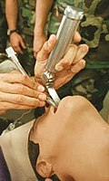
Intubation using direct laryngoscopy

Videolaryngoscope

Videolaryngoscope


Risk in difficult intubation[edit | edit source]

Part of the pre-operative evaluation by the anesthesiologist is also an assessment of the risk of difficult intubation. One of the basic predictors is the Mallampati classification, which describes the clarity of the throat after maximum opening of the mouth with tongue sticking out. The investigation is burdened with a high false positive rate.
During direct laryngoscopy, the finding is described according to Cormack and Lehane based on visible structures.
There are several other methods of assessing the risk of difficult intubation, for example, the ability to open the mouth, the shape of the chin, visible injuries, etc.
Choice of cannula[edit | edit source]

Choosing the right size and type of tube is crucial to the course of the entire operation.
- Size of endotracheal tubes according to internal diameter (mm):
- with short-term anesthesia adult men 7.5-8; women 7–7.5;
- in long-term ventilated patients, on average 0.5 mm larger (though greater discomfort after extubation and greater damage to soft tissues, but better for suctioning, bronchoscopy and improving the process of weaning from UPV due to less resistance);
- we can roughly estimate based on the size (diameter) of the patient's little finger (especially in children);
- calculation for children (age in years: 4) + 4;
- we usually use smaller caliber cannulas for nasotracheal intubation.
- with short-term anesthesia adult men 7.5-8; women 7–7.5;
- Types of tubes (for guidance only, not all types are listed):
- classic Magilas tube (standard),
- flexible Woodbridge tube – use when the patient is in the prone position, procedures on the neck or those where a rigid tube would interfere with the operator,
- tubes for one-sided intubation (Mallinckrodt, Singellumen tube) – use in procedures on the lungs and the need to ventilate only one lung,
- cannulas without an inflatable sealing cuff – most often used in children, they reduce the risk of subglottic stenoses.
Procedure[edit | edit source]
For standard uncomplicated orotracheal intubation with direct laryngoscopy in adults, we proceed as follows:
- Preparation of material, connection of monitoring.
- Positioning the patient, especially placing the head in the so-called "sniffing position".
- Sufficient preoxygenation of the patient.
- Podání analgetik a anestetik (hypnotic, opiate).
- After the onset of the effect of anesthetics, the patient is breathed through a mask and, if it is possible to breathe, administer myorelaxants. (However, sometimes the patient can breathe only after myorelaxation)
- After the onset of relaxation, we open the patient's mouth.
- We insert the laryngoscope between the root of the tongue and the epiglottis and by pulling upwards and forwards (Caution: NOT PRYING!) we get a view of the vocal cords.
- With the right hand, while constantly checking the vocal cords, we insert the tube into the glottis.
- Tube cuff inflation.
- Standard insertion depth for men is 23 cm, for women 21 cm from the front incisors. For children, we calculate (age in years : 2) + 12.
- Further adjustment of the insertion depth can be made according to the listening findings.
- We connect the tube to the anesthesia machine and verify the correct position of the tube.
Signs of successful intubation[edit | edit source]
- Certain Signs:
- direct laryngoscopy and visual inspection of the passage of the tube through the glottis,
- capnometry (CO2),
- bronchoscopy.
- Uncertain Signs:
- chest excursion,
- auscultation,
- constant pulse oximetry value.
Complications of intubation[edit | edit source]
- Mechanical damage – mucous membrane, teeth, trachea, esophagus, vocal cords;
- stimulation of reflexes – sympathotonic, vagal;
- unilateral intubation (more often right-sided);
- tube obstruction;
- bleeding;
- pressure ulcers;
- vocal cord paresis.
Difficult intubation[edit | edit source]
This condition is defined as the inability of an experienced physician to insert an intubation cannula after 3 attempts at direct laryngoscopy or the effort takes more than 10 minutes. Recommended procedures have been developed for further action.
Video[edit | edit source]
References[edit | edit source]
Related Articles[edit | edit source]
- Respiratory protection • Respiratory protection (half heels)
- Intubation (pediatrics)
- Crush Introduction to Anesthesia
External links[edit | edit source]
- Advanced emergency resuscitation – multimedia educational program, video demonstration of intubation (Clinic of Anesthesiology and Resuscitation FNKV)
- http://public.fnol.cz/www/urgent/Konference%202006/ODUM1/11_Techn_DC.pdf Brtníková Věra, Sedlák Ctirad – Techniques for securing airways KAR FN Olomouc
- Jan Bruthans – Ensuring airways KARIM 1.LF UK
- Akutne.cz: Possibilities of ensuring airway patency by general practitioners
References[edit | edit source]
- ŠEVČÍK, Pavel, et al. Intensive care medicine. 3. edition. Galen, 2014. 1195 pp. pp. 69–71. ISBN 978-80-7492-066-0.
- OREBAUGH, Steven, et al. Direct laryngoscopy and endotracheal intubation in adults [online]. UpToDate, The last revision 2020-04-29, [cit. 05/22/2020]. <https://www.uptodate.com/contents/direct-laryngoscopy-and-endotracheal-intubation-in-adults>.
- KRETZ, Franz-Josef – TEUFEL, Frank. Anästhesie und Intensivmedizin. 1. edition. Heidelberg : Springer, 2006. 695 pp. ISBN 3-540-62739-1.
- HECK, Michael – FRESENIUS, Michael. Repetitorium of Anaesthesiology. 5. edition. Heidelberg : Springer, 2007. 642 pp. ISBN 978-3-540-46575-1.