Efflorescence

Eflorescence is a cutaneous manifestation of the disease. Individual efflorescences can merge into diseased deposits or areas. The set of efflorescences is called a rash. If the rash is on the mucosa, it is called an enanthema. Primary efflorescence is the primary manifestation of the disease. Secondary arises from the development of primary efflorescence or as a secondary manifestation on deposits or surfaces.
For efflorescences we evaluate:
- localization,
- size,
- face,
- color,
- surface,
- borders,
- surroundings.
Primary efflorescences[edit | edit source]

The macula (spot) is a small surface change in color caused mainly by a change in blood flow, the number of blood vessels or pigment.
- freckles, vitiligo, some drug rashes

The papule (bud) is a small skin prominence up to 1 cm.
- psoriasis, warts
- papulopustular − a bud containing a cavity filled with pus (acne)
- papulovezicular − a bud with a tube with a clear content
Tuber (bump) is a papule of larger dimensions (over 1 cm).
- in furuncle, spinalioma, lipoma
Pomphus (urticaria bud, urtica) is a bud caused by corium edema, lasts short, usually itchy.
- utricaria (hives), insect bites (mosquito)

The vesicle (blister) is a cavity filled with clear contents.
- subcorneal − impetigo
- intraepidermal − pemphigus vulgaris, eczema, herpes simplex
- subepidermal − bullous pemphigoid, dermatitis herpetiformis (Duhring)
The bulla is larger , arises, for example, in Lyell's syndrome, bullous form of impetigo.
The pustule is a cavity filled with pus. More often, however, it occurs secondarily by turbidity of the vesicle contents.
- primary − folliculitis, psoriasis pustulosa

The nodule is a bounded round formation that results from changes in the corium or subcutaneous tissue. It can be palpable below the surface or it can be raised.
- For example: erythema nodosum, lipoma.
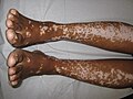
Vitiligo − merging macules

Warts − papular lesions on the index finger
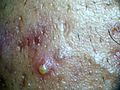
Pustula


Secondary efflorescences[edit | edit source]

Squama (scale) a scale or plate-like structure, dry or greasy masses of keratin
- pityriaziform − bran-like scales (pityriasis versicolor)
- lamellar− flaky(chronic eczema)
- exfoliative− bar-shaped (scarlatina)
Crust (scab) is formed by drying of tissue fluid (yellow color), pus (yellow-green color) or blood (brown to black color).
- crustosquama − scale permeated with liquid and dried
- rupee − terraced
Eschara (plaque) is caused by the death of the skin, the dead tissue is initially whitish, later gray-brown to black, bordered by demarcation inflammation, leaves behind an ulcer, heals with a scar.
- eg burns, trophic circulatory disorders
Rhagade (fissure) is a cleft disruption of skin continuity, formed by rupture only of the epidermis; if it gets under the epidermis, it bleeds - fissura (crack).
- in case of impaired skin elasticity (edema, infiltrate, hyperkeratosis ...), in places of mechanical stress
Erosion − a superficial defect affecting only the epidermis or mucosa, wet when the stratum spinosum is exposed, rupture of the papillae will cause fine bleeding.
Excoriation is caused by mechanical scraping (in pruritus).
Ulcus (ulcer) is a deep skin defect, it always reaches the corium or deeper, we describe the size and shape, the bottom (clean, coated; fine, bumpy, ...)
Aphtha is an efflorescence occurring only in the oral cavity, it is an erosion covered by fibrin, bounded by a reddened rim.

Ulcus cruris
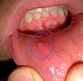
Aphtha


Other terms used to describe skin changes[edit | edit source]
Erythema − red skin color.
Edema – diffuse swelling of the skin.
Madidatio (maceration) – wetting of tissue fluid; softering and turning white due to beeing constantly wet
Cicatrix (scar) − is formed by the transformation of granulation tissue (loss of fibroblasts and capillaries), fresh is pink, old is whitish. The scar can be atrophic, hypertrophic, keloid.
Lichenification is thickening of the skin.
Links[edit | edit source]
Related articles[edit | edit source]
- Infectious exanthematous diseases in childhood
- Configuration of efflorescences
- Localization of efflorescences
Refferences[edit | edit source]
- ŠTORK, Jiří, et al. Dermatovenerologie. 1. vydání. Praha : Galén, Karolinum, 2008. ISBN 978-80-7262-371-6.
- JIRÁSKOVÁ, Milena. Dermatovenerologie pro stomatology. 1. vydání. Praha : Professional Publishing, 2001. ISBN 80-86419-07-X.
- Kangle S, Amladi S, Sawant S. Scaly signs in dermatology. Indian J Dermatol Venereol Leprol [serial online] 2006 [cited 2020 Nov 24];72:161-4. Available from: https://www.ijdvl.com/text.asp?2006/72/2/161/25653