Development of body cavities and mesenteries
From WikiLectures
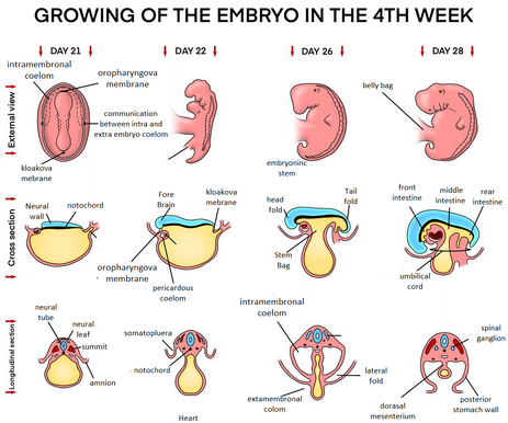
 At the beginning of the 4th week of embryonic development, the ``intraembryonic coelom takes the form of a horseshoe-shaped curved cavity in the cardiogenic and lateral mesoderm. The anterior part represents the future pericardial cavity and its arms the pleural and the peritoneal cavity. Both arms open into the extraembryonic coelom in their distal sections. In the embryos of lower animals, excretory products are temporarily stored throughout, in humans it provides space for the displacement of organs.
Body cavities of the embryo[edit | edit source]
- The cavities are lined with a parietal wall lined with mesothelium (most of the future parietal layer) derived from somatic mesoderm,
- and then through the visceral wall arising from the splanchnic mesoderm,
- the peritoneal cavity is connected to the extraembryonic coelom in the region of the umbilical cord,
- loses its connection in the 10th week, when the intestines return from the umbilical cord back to the abdominal cavity (see below),
- after the formation of folds is completed, the caudal section of the foregut, midgut and hindgut are attached to the back wall by means of the dorsal mesentery.
Mesentery[edit | edit source]
- It arises as an extension of the visceral sheet of the peritoneum covering the organs,
- dorsal and ventral mesenteries temporarily separate the peritoneal cavity into right and left, but the ventral mesentery soon disappears,
- arteries supplying the primitive intestine,
- anterior intestine – truncus coeliacus,
- middle intestine – superior mesenteric artery,
- posterior intestine – arteria mesenterica inferior.
Division of embryonic coelom[edit | edit source]
- After the embryo curls, the flat coelom cavity twists with its pericardial part down, then it is deposited on the sides of the foregut,
- The septum transversum, which formed the mesenchymal mass in front of the germinal target, reaches ventrally and caudally from the pericardial coelom and begins to separate the thoracic region from the abdomen (it later forms the center of the tendineum of the diaphragm),
- at the same time, septa are formed in both pericardial channels, which begin to separate the individual serous cavities,
- it is the result of ingrowth of bronchial buds into the lumen of the pericardioperitoneal channels,
- piles,
- plicae pleuropericardiales – located above the developing lungs,
- plicae pleuroperitoneales – below the base of the lungs.
Pleuropericardial membranes[edit | edit source]
- Plicae pleuroperiacardiacae – separate the thoracic and pericardial cavity,
- pleuses contain common cardiac veins (venae cardinales communes), which drain blood from the primitive venous system into the sinus venosus,
- bronchial buds grow into the pleural cavity, which expands ventrally and separates the mesenchyme into two layers,
- the outer layer that will form the chest wall,
- the inner layer which envelops the pericardial cavity and which will form the fibrous pericardium,
- due to the growth of the cardiac veins, the descent of the heart and with the expansion of the pleural cavity, the pleuropericardial membranes turn into serous duplicatures that depart from the lateral surface of the chest,
- by the seventh week, they fuse with the mesenchyme in front of the esophagus and thus separate the pericardial space from the pleural cavities.
Pleuroperitoneal membranes[edit | edit source]
- These cysts gradually grow into the lumen of the pericardioperitoneal channels, then become relatively thin and turn into pleuroperitoneal membranes,
- gradually separates the thoracic and abdominal cavity,
- arise from the expansion of the developing lungs and pleural cavities that penetrate the body wall,
- they grow from the dorsolateral wall and are attached to the abdominal wall, they initially protrude into the cavity,
- during the 6th week, the membranes expand ventromedially and merge with the dorsal mesentery of the esophagus and the septum transversum,
- closure of the pleuroperitoneal passage is accompanied by migration of myoblasts,
- the opening on the right side closes earlier than on the left (probably due to the larger size of the liver).
Links[edit | edit source]
Related Articles[edit | edit source]
- Stages of embryo and fetal development: First week of human development • Second week of human development • Third week of human development • Fourth to eighth week of intrauterine development
References[edit | edit source]
- MOORE, Keith L. – PERSAUD, T.V.N.. The Birth of Man. 1. edition. ISV, 2002. 564 pp. ISBN 80-85866-94-3.
