Brain
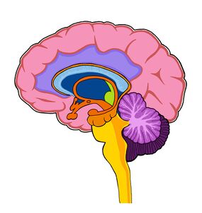
The central nervous system is anatomically divided into the brain (encephalon, cerebrum) and the spinal cord. The brain is the part stored in the cranial cavity that protects it from injury. Anatomically, the brain consists of several parts that arose in embryogenesis from the neural tube. The spinal cord is followed by the medulla oblongata, Varol's bridge (pons Varoli), mesencephalon, diencephalon and telencephalon. The cerebellum sits dorsally on the brainstem (spinal cord + pons + mesencephalon). Its structures are separated only anatomically, functionally there is a wide constellation of pathways and connections of various importance between them.
In the brain, the so-called gray and white brain matter are distinguished. The gray matter, which consists mainly of the nerve cell bodies of neurons, covers the surface of the cerebrum as the cerebral cortex and forms the so-called nuclei, which are stored inside the other sections of the brain. The white matter is made up of nerve cell axons. Inside the brain there are four brain chambers, between which and the space around the brain (encephalon, cerebrum) and spinal cord and meninges circulates cerebrospinal fluid. Brain functions are very complex.
Brain stem[edit | edit source]
The brainstem connects rostrally to the spinal cord (medulla spinalis) and consists of the medulla oblongata, Varoli's bridge (pons Varoli) and the midbrain (mesencephalon). The brain stem has a number of structures in common, and a number of pathways or nuclei run through the entire stem and do not respect its division into the medulla oblongata, pons and midbrain at all.
Medulla oblongata[edit | edit source]
The elongated spinal cord (medulla oblongata) is a continuation of the spinal cord in the rostral direction and, as its most caudal part, already belongs to the brain. The dividing line between the spinal cord and the spinal cord is the decussatio pyramidum. The border of the medulla oblongata and Varola's bridge is the sulcus bulbopontinus, a groove running horizontally at the rostral end of the medulla oblongata. Ventrally, the elongated spinal cord is arched into two parallel, longitudinal mounds - pyramides medullae oblongatae, which contain the white matter of the pyramidal pathway, i.e. tractus corticospinalis. The median anterior fissure runs between them.
Laterally, there are paired elevations, called olives, on the elongated spinal cord. Dorsal to the olive lie the pedunculi cerebellares inferiores. The pedunculi cerebellares are generally thick bundles of white matter through which the pathways connect the stem (in this case the medulla oblongata) to the cerebellum. The peduncles diverge into a V-shape, and between them is spread the velum medullare inferius, which is a thin plate, an outgrowth of the ependyma. The free end of the vela is joined by the tela choroidea ventriculi quartii, a fibrous plate containing the choroid plexus, which forms the cerebrospinal fluid, here to IV. cerebral ventricles.
Tela choroidea ventriculi IV. it is not whole, it contains several holes. They are apertura median ventriculi quarti (foramen Magendi) and paired aperturae laterales ventriculi quartii (foramina Luschkae). These openings form a communication between the ventricular system and the subarachnoid space of the brain and allow the circulation of cerebrospinal fluid. The dorsal surface of the elongated spinal cord is also arched into two bumps - tuberculum gracile and tuberculum cuneatum. They contain the nuclei of the same name, which are the final station of the spinal fasciculus gracilis and cuneatus, which lead the main sensitive pathways of the brain. The pathways connect here and continue further into the higher levels of the brain. The canalis centralis runs through the oblongata, which opens cranially into the IV. cerebral ventricles. The following cranial nerves depart from the medulla oblongata – IX, X, XI and XII.[1]
Pons Varoli[edit | edit source]
Varol's bridge is a continuation of the medulla oblongata rostral to the sulcus bulbopontinus. It forms an oval arch on the ventral side of the brainstem. It passes cranially into the midbrain. The ventral side of the bridge is smooth and convex. The sulcus basilaris, formed by the course of the artery of the same name, leads through the medium. Laterally, the bridge passes freely in the pedunculi cerebellares medii, analogous to the pedunculi cerebellares inferiores of the medulla oblongata. Like the medulla oblongata, the pons is also related to the cranial nerves, which have their nuclei here. Nerves V, VI, VII and VIII depart from the pons.
Mesencephalon[edit | edit source]
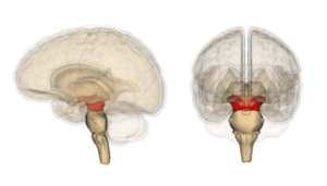
The middle brain (mesencephalon) is the most rostral part of the brainstem, it connects to Varol's bridge.
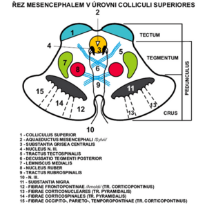
It connects the rhombencephalon to the diencephalon. It measures about 2cm in length. Practically all of it is covered by the hemispheres of the hindbrain, only its ventral part is visible as the so-called crura cerebri (partes anteriores pedunculi cerebri) – massive trunks containing white matter. The aquaeductus mesencephali (Sylvii) runs through the middle brain – a narrow channel, carrying the cerebrospinal fluid, after the distance from IV. chambers.
Aquaeductus m. is lined with a layer of gray matter substantia grisea centralis.
The midbrain can be divided into several parts - tectum mesencephali and pedunculus cerebri (which consists of tegmentum mesencephali and crura cerebri).
The pedunculus cerebri is ventral to the aquaeductus m.
Crura cerebri (partes anteriores pedunculi cerebri) - also part of pedunculus cerebri. They are lateral and sink under the optic tract into the base of the brain.
Between the two trunks is the fossa interpeduncularis. Its surface is perforated by the course of a series of vessels, therefore it is called substantia perforanta posterior (interpeduncularis). Be careful not to confuse the substantia perforanta anterior, which is part of the hindbrain!
In the fossa interpeduncularis, medially from the pedunculi, the third oculomotor nerve emerges.
Ventral to the fossa interpeduncularis are the corpora mamillaria, which already belong to the diencephalon. These cranial nerves depart from the midbrain – III, IV (II runs here already as a pathway, not the optic nerve). The cranial ends of the reticular formation also extend here.
The tectum lies dorsal to the aquaeductus mesencephali. It contains two pairs of bumps - colliculi superiores et inferiores.
They are involved in the visual and auditory pathways and continue as the brachium colliculi superioris et inferioris to the corpus geniculatum laterale and mediale diencephala.
Caudal to the colliculi extend the pedunculi cerbellares superiores, another in the series of cerebellar tracts. As with the pedunculi cerebellares inferiores, the velum, more precisely, the velum medullare superius, forming the cranial part of the IV ceiling, is opened here. chambers. Cranial to the tectum is the area pretectalis, which already belongs to the diencephalon
Nuclei of the tectum mesencephali
- predominantly sensitive pathways (sight and hearing)
a) Nc. colliculi superioris - fibers from the retina of the eye as well as motor and somatosensitive
b) Nc. colliculi inferioris - auditory cortex center
c) Nc. posterior commissures
d) Ncc. pretectales - nc. optic tract
Tegmentum (partes posteriores pedunculi cerebri) – ventral to tecta, part of pedunculus cerebri.
The boundary between the crura and the tegmentum is formed by the substantia nigra.
Substantia nigra - pigment in the perikarya, the outer part goes to the crura and has a net-like appearance (pars reticularis) and the inner part goes to the tegmentum (pars compacta) produces dopamine.
It contains a number of important pathways and nuclei. On the side, a gentle elevation is visible - the trigonum lemnisci, where the lemniscus medialis runs.
Nuclei of the tegmentum mesencephali
- mainly motor paths
a) Nc. ruber - oval, large, reddish, between substantia nigra and aquaeductus, regulation of limb movements
b) Nc. nervi oculomotorii - next to the aquaeductus, a set of several nuclei, sends somatomotor fibers to the oculomotor nerve, which innervate 4 of the 6 oculomotor muscles, controls the movements of the eyeball
c) Nc. accessorius n. oculomotorii - dorsal from nc. nervi oculomotorii, visceromotor parasympathetic fibers, ciliaris muscle and sphincter pupillae, movements of the eyeball
d) Nc. interstitialis Cajal - small, dorsal from nc. nervi oculomotorii, related to the gray matter of the substantia grisea centralis and to the rostral part of the formatio reticularis, part of the fasciculus longitudinalis medialis
e) Nc. Darkshevics - small fibers from the cerebellum, cortex of the telencephalon, vestibular nuclei of the rhombencephalon
f) Nc. nervi trochlearis - small, next to the substantia grisea centralis, caudal from n. III, somatomotor fibers innervate the obliquus superior bulbi muscle
g) Nc. mesencephalicus nervi trigemini - long and slender, it goes here from the rhombencephalus, laterally from the aquaeductus, sensitive muscle and joint receptors of the masticatory muscles, artic. temporomandibularis and oculomotor muscles
Pathways of the mesencephalon:
- afferent to ncc. collicules superiores from the retina of the eye, from the spinal cord, from the rhombencephalon, from the visual cortex
- to colliculi inferioris from ncc. cochlear
Cerebellum[edit | edit source]
The cerebellum is located in the posterior fossa of the skull, dorsal to the medulla oblongata and the brainstem.
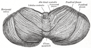
It is a rounded dorsally arched formation. A round, longitudinal, narrow middle band, separated by sagittal depressions from the lateral parts = vermis cerebelli (cerebral worm). Hemisphaeria cerebelli: 2 lateral, larger, symmetrically constructed hemispheres. The cranial surface is flatter, contact with the roof-like duplication of the dura mater (tentorium cerebelli). Dorsal and caudal surfaces arched; stored in the pits of the occipital bone under the transverse arms of the eminentia cruciformis (fossae occipitales cerebellares). The falx cerebri extends between the hemispheres of the cerebellum (from the crista occipitalis interna).
3 pairs of peduncles, pedunculi cerebellares, enter the cerebellum from the brainstem:
- Inferiores (corpora restiformia) – connect the oblongata with the cerebellum; they line the caudal part of the fossa rhomboidea.
- Media (pontini; brachia pontis) – connect the pons with the cerebellum; they border the fossa rhomboidea.
- Superiores (brachia conjunctiva) – connect the tegmentum mesencephali with the cerebellum; they border the rostral part of the fossa rhomboidea.
All peduncles contain pathways going to and from the cerebellum. Between the pedunculi cerebellares superiores, the velum medullare superius (craniale) is opened - the front part of the ceiling IV. ventricles, drawn up into a peak called the fastigium.
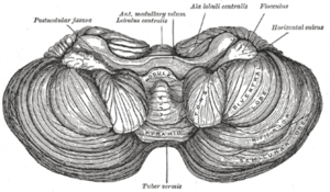
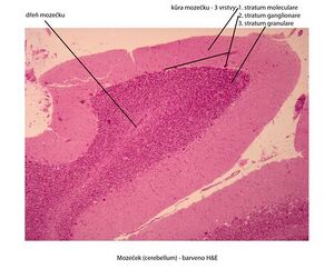
On the surface of the cerebellum, numerous transverse furrows - they separate individual sections on the vermis and hemispheres = fissurae cerebelli. The largest and deepest fissures separate 3 hl. sections: lobi cerebelli. Smaller fissures further divide these lobes into lobules: symmetrically placed on the hemispheres; correspond to an odd section on the vermis. The smallest fissures separate the parallel strips of the surface of the cerebellum = folia cerebelli. The surface is covered by a continuous gray matter: cortex cerebelli. Fissura prima - from the center to both sides, fissura horizontalis - dorsal pole, fissura posterolateralis - separates nodulus and flocculus.
Inside the cerebellum is the white matter, corpus medullare. It extends in the form of plates as laminae albae into the folia of the cerebellum. On a sagittal section, the vermis forms a tree-like pattern (arbor vitae, tree of life).
Paired clusters of gray matter are stored in the white matter - nuclei cerebelli:
- Ncl. dentatus – the largest of the cerebellar nuclei. Two parts: dorsomedial (paleocerebellar) with fibers going to the ncl. ruber and ventromedial (neocerebellar) with fibers going to the thalamus. The appearance of a wrinkled pouch with an opening ventromedially against the mesencephalon. Opening of the pouch = hilum (hilus) nuclei dentati. Thence the path contained in the pedunculus cerebellaris superior.
- Ncl. emboliformis – an elongated small core, the shape of a blood clot. Filed sagittally at the hilum of the ncl. dentatus.
- Ncl. globosus – in pairs, placed medially from ncl. emboliformis. From several small spherical formations of gray matter.
- Ncl. fastigii – paired, placed most medially at the fastigii, near the midline.
Ncl. emboliformis, globosus and fastigii efferent to the ncl. ruber, reticular nuclei, mesencephalon, pontus and oblongata. All the cerebellar nuclei are the starting point of the pathways coming out of the cerebellum - through them the cerebellum is involved in the movement control system.
The nuclei contain the bodies of multipolar neurons on which the axons of the Purkinje cells terminate.
Morphological division of the cerebellum[edit | edit source]
It is divided into 3 lobes by transverse grooves. In each lobe, the lobules are separated by smaller grooves (on the vermis and hemispheres). Morphological division enables a topographical orientation to the cerebellum, but does not correspond to the developmental and functional division.
Description of formations[edit | edit source]
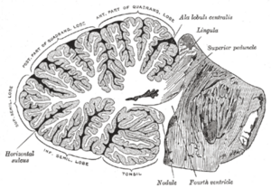
Top surface (front to back):
Vermis (lobus cerebelli anterior)
- Lingula cerebelli – 1 to several foils resting on the velum medullare superius.
- Lobulus centralis – a square group of foils in the front incisor. (fissula precentralis)
- Monticulus – the larger part of the upper surface of the vermis, hump-shaped; it is divided into culmen and declive (fissura prima) by a transverse groove
- Folium vermis – the only folium at the incisura cerebelli posterior
Hemispheres
- Vinculum lingulae cerebelli – narrow white band.
- Ala lobuli centrales – a triangular group of folia in the anterior incisor.
- Lobulus quadrangularis – divided by a transverse groove into pars sup. et inf. (pars inf. otherwise also lobulus simplex).
- Lobulus semilunaris vulture. – crescent shaped.
The lower surface (it is separated from the upper surface by the fissura horizontalis cerebelli):
Vermis (lobus cerebelli posterior)
- Tuber vermis – protrudes into the incisura cerebelli post. (horizontal fissure)
- Pyramis vermis – the widest part of the worm ( fissura prepyramidalis )
- Uvula vermis – elongated elevation of several foils ( fissura secunda )
- Nodulus vermis – attached to the uvula, rests on the velum medullare inf.
Hemispheres
- Lobulus semilunaris inf – lobulus gracilis joins it.
- Lobulus biventer – bulging.
- Tonsila cererebelli – considerably convex groups of horseshoe-shaped folia.
- Flocculus – a stalked group of foils with a curly edge; a rudimentary paraflocculus attaches at the posterior margin.
Division of departments according to development relationships[edit | edit source]
Vestibular cerebellum – the oldest part, the basis of development are the vestibular pathways. It consists of: flocculus, lingula and nodulus.
Spinal cerebellum - the basis of development are the spinocerebellar pathways, it divides the vestibular cerebellum into the front (lingua) and back (nodulus and flocculus) parts. It consists in front: lobulus centralis, culmen, lobulus quadrangularis superior and behind: pyramis, uvula, paraflocculus.
Cerebral cerebellum – develops by afferent from the cortex, through the pontocerebellar pathway. It arises in the middle of the older parts of the cerebellum collectively referred to as the palaeocerebellum. It is then called the neocerebellum (seu lobus medius).
The furrowing of the cerebellum happens gradually:
The sulcus primarius (fissura prima) forms the earliest. It separates a part called the anterior lobe (rostralis). This includes the lingua and the anterior part of the spinal cerebellum. The following groove - fissura praepyramidalis defines the lobus medius and lobus caudalis in the area of the vermis. Fissura nodulouvularis - border between lobus caudalis and pars nodulofloccularis.
Division including antaomical and develompental characteristics[edit | edit source]
- Lobus rostralis (anterior) – the anterior rudiment of the vestibular cerebellum and the anterior spinal cerebellum.
- Lobus medius – the largest part, includes the cerebral cerebellum.
- Lobus caudalis (posterior) – posterior spinal cerebellum.
- Lobus nodulofloccularis – part of the vestibular cerebellum not included in the lobus rostralis.
Functional connectivity of the cerebellum[edit | edit source]
Supply pathways via the pedunculi cerebellares inferiores, medii, superiores to the cerebellar cortex. The exit of fibers from the cerebellar cortex ends in the cerebellar nuclei.
Cerebellar nuclei send axons to the gray matter of the brain stem (mainly to the reticular formation, ncl. ruber, to the thalamus). Pathways to the spinal cord originate from the gray matter, which influence cells that send their axons as motor fibers to the skeletal muscles. It directs and controls movement activities and muscle tone, when the vermis participates in the coordination of the trunk muscles and the hemispheres of the muscles of the ipsilateral limbs.
Cerebellar pathways[edit | edit source]
Afferent pathways mainly go to the cerebellar cortex. Efferentation begins with Purkinje cells (1st neuron) and continues after switching in the cerebellar nuclei (2nd neuron) in the centrifugal pathways going outside the cerebellum.
Vestibulocerebellum - lobus flocculonodularis and lingula vermis are connected to the vestibular nuclei of the rhombencephalon, from the ncc. vestibular and ncc colliculi superiores and visual cortical areas, maintaining body balance, spatial orientation.
Spinocerebellum - lobus cerebelli posterior, anterior except for the rostral section of the lingula connects to the spinal cord, af. fibers go from the trigeminal nerve, auditory and visual structures of the CNS and ef. they go to the motor nuclei of the brainstem, their function is motor coordination and they respond to proprioceptive information.
Cerebrocerebellum - through nuclei pontis connected with cerebral cortex, af. fibers from the cerebral cortex via the ncc. pontis, ef. they are to the motor thalamus (nc. ventralis anterior and lateralis) and to the cerebral cortex, the function is the coordination and timing of muscle movements.
A) Tracks of the partis nodulofloccularis:
- Afferentation from ncll. vestibulares as tr. vestibulocerebellares.
- Efferentation goes to Deiters nucleus, according to its origin as tr. nodulovestibularis and flocculovestibularis. From ncl. fastigii goes separately tr. fastigiovestibularis (Russell's bundle) along the pedunculus cerebellaris inf. to the Deiters core.
B) Pathways lobi rostralis et lobi caudalis:
Afferent pathways:
- Tr. spinocerebellaris post.: Stiling-Clark nucleus - pedunculus cerebellaris inf. - cerebellar cortex (and cerebellar nuclei).
- Tr. spinicerebellaris ant. (Gowersi): crossing in the spinal cord - lateral spinal cord (ventral from tr. spinocerebelaris post) - through the pedunculi cerebellares superiores to the cerebellum.
- Tr. bulbocerebellares: from the nuclei of the posterior cords of the spinal cord - uncrossed as fibrae arcuatae externae dorsales or crossed as fibrae arcuatae externae ventrales and fibrae arcuatae internae - through the pedunculus cerebellaris inf.
- Tr. nucleocerebellares: from the nuclei of sensitive cranial nerves.
- Tr. olivocerebellares: from the major olive (older part) and minor olive.
- Tr. tectocerebellaris: from the gray matter under the colliculi superiores - velum medullare superius - cortex vermis superior.
- Tr. reticulocerebelaris: from the nuclei of the lateral nuclei of the RF - pedunculus cerebelaris inf. - vermis - to the ipsilateral hemisphere.
- Tr. rubrocerebellaris: after crossing tr. rubrospinalis branches off into - pedunculus cerebellaris sup.
Efferent pathways:
- Tr. cerebellotegmentalis (dentatotegmentalis): from the cerebellar nuclei (mainly incl. dentatus) - pedunculus cerebellaris sup. - FR nuclei of the pontine and mesencephalon.
- Tr. cerebellorubralis (dentatorubralis): via pedunculi cerebelli sup. to ncl. ruber (then using tr. rubrospinalis and rubroolivaris for olive).
- Tr. cerebelloolivaris: via pedunculi cerebelli inf. - contralaterally to the main olive (older part) and secondary olive.
- Tr. cerebellotectalis: via pedunculi cerebelli sup.
- Tr. cerebellothalamicus (embolothalamicus): through the central nuclei of the thalamus to the striatum
C) Lobi media pathways
Afferent pathways:
- Tr. pontocerebellares: from ncll. pontis, where they cross - pedunculi cerebelli medii - cortex of the cerebellum; the pathway is a continuation of the corticopontine pathway (tr. frontopontinus et tr. occipitotemporopontinus) and cross-connects the hemispheres of the forebrain and cerebellum.
- Tr. olivocerebellares: as in the orbits of the lobi rostralis et caudalis, but it originates from the neooliva (younger part of the ncl. olivaris).
- Tr. corticocerebellares: from the motor area of the frontal lobe - pedunculi cerebelli inf. - to the ipsilateral hemisphere of the cerebellum.
Efferent pathways:
- Tr. cerebellorubrales: conduction as in the same-named pathway from the previous group of pathways.
- Tr. cerebrothalamici: from ncl. dentatus to the ventrolateral nuclei of the thalamus (further as tr. thalamocorticalis to areas 4 and 6).
Diencephalon[edit | edit source]
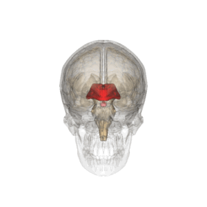
Diencephalon also called midbrain consists of 5 functionally and morphologically distinct parts. Dorsoventrally they are: epithalamus, thalamus, metathalamus, subthalamus and hypothalamus.
Anatomy[edit | edit source]
The midbrain connects to the upper end of the brain stem. It is located between the hemispheres of the terminal brain, thus it is not well visible. The only visible structure lies on the ventral surface of the brain, and that is the hypothalamus. The posterior border is formed by the upper end of the interpeduncular fossa, or two bumps, the corpora mamillaria. It ends in the area of the optic chiasm.
The diencephalon is formed by further brain development of the anterior cerebral sac (prosencephalon), in which the original division to allar a basal discs is evident. The thalamus (sensitive structure) and basal hypothalamus (visceromotor structure) develop from the alar plates.
Description[edit | edit source]
The most conspicuous part of the midbrain are the two arches, which are the thalami that form the lateral wall of the '''IIIrd ventricle'''. Furthermore the choroidal fibrous bodies of the IIIrd ventricle form the ceiling of the 3rd ventricle. The place of attachement of the choroidal body is called taenia thalami.
The diencephalon contains the 3rd cerebral ventrical, which is the continuations of the aquaeductus mesencephali, coming from the IV.th cerberal ventricle. It then flows into the intraventricular foramina, through which it enters the lateral ventricles (between the hemispheres of the terminal brain).
The medial wall of the diencephalon (side walls of the III. ventricle) is divided by a paired groove - sulcus hypothalamicus (corresponds to sulcus limitans of the neural tube). This structure divides the diencephalon into dorsal and ventral parts. The dorsal part includes the thalamus, metathalamus and epithalamus, which are mainly sensitive (sensory). The ventral part includes the subthalamus and hypothalamus, whose functions are mainly motoric.
Epithalamus[edit | edit source]
Is the dorsocaudal part of the diencephalon, which consists of habenular nuclei and pineal body. The habenular nuclei are contained in the habenular trigone, which is formed by the extension of the bundle of white matter (stria medullaris thalami). Both trigones together form the habenula, within which fibers of the of the stria medullaris thalami cross. In this area of crossing the pineal body (epiphysis) extends from the epithalamus.
- Nuclei
- Inside the habenula there are habenular nculei (nucleus habenularis medialis et lateralis). Their activity is somatomotor and visceromotor, allowing reactions of olfactory and limbic arousal. Habenula is a functional part of the limbic system.
- Tracts
- Commissura posterior connects posterior thalamic nuclei, colliculi superiores and pretectal nuclei of both sides. It contains fibers emerging from the ncl. interstitialis, ncl. Darkshevichi, pretectal nuclei and a part of habenulotektal fibers.
Thalamus[edit | edit source]
Thalamus presents a paired part of the diencephalon and it is oval in shape
The anterior part narrows to the anterior tubercule and posterior rounded part is referred to as the pulvinar. The 2 parts of the thalamus are
joined to each other through the interthalamic adhesion.
Metathalamus[edit | edit source]
The metathalmus occipitally follows the thalamus. It consists of the lateral geniculate body, which is located under the pulvinare and mediale. The metathalamus is involved in the visual pathway and auditory pathway, receiving signals from the mesencephalon.
- Nuclei
- Ncl. corporis geniculati lateralis belongs to the visual pathway and ncl. corporis geniculati medialis belongs to the auditory pathway.
Subthalamus[edit | edit source]
Lies ventrally from the thalamus and laterally from the hypothalamus.
Hypothalamus[edit | edit source]
A small part of the diencephalon is found under the thalamus. Rostrally it reaches up to the lamina terminalis and caudally to the posterior margin of the mamillary bodies. It lies laterally to the III. cerebral venrticle and medially to capsula interna. The infundibulum protrudes on the base of the hypothalamus and continues as a stalk on which the hypophysis (pituitary gland) hangs.
Hypothalamus serves as the highest viscermotoric center in the human body. Further on it is the center of activities of the autonomic nervous system. Its functions include alsoendocrine activities.
Telencephalon[edit | edit source]
In humans, the largest part is the hindbrain (telencephalon). It is covered with cerebral grooves (sulci cerebrales) and turns (gyri cerebrales). Greater grooves separate the 5 telencephalic lobes:
- frontal;
- parietal;
- occipital;
- temporal;
- insular.
Cortex cerebralis[edit | edit source]
The telencephalon is covered by the cerebral cortex. Wrinkling allows the area of the cerebral cortex to increase several times. In simple terms, it can be said that the cerebral cortex is co-responsible for consciousness, it plays an essential role in perception, thinking, memory, mental abilities, and in the initiation of free movements. The locations of some of these functions are known, e.g. speech center, visual center, etc.
White matter of the telencephalon[edit | edit source]
- the substantia alba fills the inner spaces between the nuclei and the lateral chambers under the cortex
- it consists of axons and dendrites with glial cells, blood vessels and other parts of the nervous system, we call it as neuropil
- nerve fibers of the white matter are divided into 3 groups:
- Association fibers - connect different places of the same hemisphere to each other
a) short - fibrae arcuatae cerebri - connect the coils
b) long - they connect different lobes of the same hemisphere, e.g. a bundle of fibers runs longitudinally from front to back under the cortex of the gyrus cinguli - cingulum, connects the frontal, occipital and temporal lobes, then fasciculus longitudinalis superior, fasciculus longitudinalis inferior and fasciculus uncinatus - connects frontal with temporal lobe, fasciculi occipitales verticales
- Commissural pathways - fibers that run across and connect identical and different places of the right and left hemispheres
a) commissura anterior - in the front wall of the 3rd ventricle near the lamina terminalis, fibers connect the temporal lobes and areas of the olfactory cortex
b) commissura hippocampi - connects the right and left gyrus parahippocampalis
corpus callosum - the most powerful commissure of the telencephalon, starts from the lamina terminalis as a rostrum, continues through the front bend of the genu into the horizontal part of the truncus and expands at the end as a splenium, on a horizontal section the fibrae corporis callosi can be seen, which form such a bent shape of the forceps minor and major at the back
- Projection fibers - long, bring information to the cortex from lower parts of the CNS or lead commands to lower parts
- they are significant at the level of the internal capsule - medial from the nucleus lentiformis, it has the crus anterius, genu capsulae internae and crus posterius
Links[edit | edit source]
Related articles[edit | edit source]
Source[edit | edit source]
- JANČÁLEK, Radim a Petr DUBOVÝ. Základy neurověd v zubním lékařství [online]. MEFANET, ©2011. Poslední revize 27.10.2011, [cit. 26.11.2011]. <http://portal.med.muni.cz/clanek-560-zaklady-neuroved-v-zubnim-lekarstvi.html>.
- VOKURKA, Martin. Velký lékařský slovník. 7. vydání. Praha : Maxdorf, 2007. ISBN 978-80-7345-130-1.
- Flash animace – e-learning 1. LF UK Praha
- ČIHÁK, Radomír. Anatomie III. 2., upr. a dopl vydání. Praha : Grada Publishing, spol. s. r. o., 2004. 673 s. ISBN 80-247-1132-X.
- ČIHÁK, Radomír a Miloš GRIM. Anatomie. 2. upr. a dopl vydání. Praha : Grada Publishing, 2004. 673 s. sv. 3. ISBN 80-247-1132-X.
- ČIHÁK, Radomír. Anatomie 3. 2. vydání. Praha : Grada, 2002. 516 s. ISBN 80-7169-970-5.


