Hypophysis
The pituitary gland (pituitary gland, lat. glandula pituitaria) is the central organ of the endocrine system. It is located behind the chiasma opticum, in the recess of the wedge bones called the Turkish saddle (sella turcica), in its deepest part, the fossa hypophysialis. It is suspended on the peduncle (infusion), which protrudes from the hypothalamus at the base of the diencephalus. It is largely superior to all other glands of the endocrine system. It's superior surface is covered by the diaphragma sellae( reflection of the dura mater).
Development of Hypophysis[edit | edit source]
It consists of two compartments, the anterior adenohypophysis and the posterior neurohypophysis. These compartments differ not only in their function, but also in embryonic origin. Adenohypophysis (Pars distalis) and intermediate lobe (Pars intermedia) originate as Rathke's bulge from the roof of the primitive oral cavity – stomodea in the third week of embryonic development. Infundibulum is the downward growth of the diencephalon that becomes the pituitary stalk and neurohypophysis. Thus, the adenohypophysis is of ectoderm origin, while the neurohypophysis is of neuroectoderm origin.
Adenohypophysis[edit | edit source]
The adenohypophyis is derived from the Rakthe's pouch( outpouching of the roof of the pharynx)
It consists of pars distalis, pars tuberalis and pars intermedia.
Pars distalis[edit | edit source]
Pars distalis is composed of beamed epithelium, sparse collagen connective tissue and blood sinusoids. It contains chromophobic and chromophilic cells. Chromophilic cells stain intensively due to the number of cytoplasmic secretory granules where hormones are stored. Chromophobic cells stain little and represent either degraded chromophilic cells or follicular cells that provide support to chromophilic cells. Chromophilic cells are further divided into acidophilic cells (stained intensively with eosin) and basophilic cells (stained with hematoxylin).
Acidophilic cells produce simple proteins and predominate in the beamed epithelium, there are two main types of cells:
- somatotropic cells − somatotropin (STH, growth hormone);mammotropic cells − prolactin (PRL).
Basophilic cells secrete glycoproteins – they are PAS positive:
- thyrotropic cells − thyrotropic hormone (TSH);
- gonadotropic cells − follicle-stimulating hormone (FSH), luteinizing hormone (LH);
- corticotropic cells − adrenocorticotropic hormone (ACTH).
Pars tuberalis[edit | edit source]
Funnel-shaped upper protuberance of pars distalis surrounding the upper scape of the infundibula. It contains mainly gonadotropic cells, they are chromophobic.
Pars intermedia[edit | edit source]
Pars intermedia is the section between the posterior and anterior lobes of the pituitary gland. There are variously sized closed follicles, so-called. Rathke's cysts, which are lined with single-layered epithelium and contain a colloid. There are also beams of chromophobic cells and also basophilic melanotropic cells secreting melanocytes stimulating hormone (MSH).
Functions of the anterior pituitary gland hormones[edit | edit source]
- Growth hormone (somatotropins): a peptide hormone, that stimulates growth via production of Insulin-like growth factor 1 (IGF-1) and increases the concentration of glucose and free fatty acids
- Prolactin (mammotrophs): Stimulates the secretion of milk from the mammary gland
- Thyroid stimulating hormone (thyrotrophs): Stimulates the production and secretion of thyroid hormone and calcitonin by the thyroid gland.
- Adrenocorticotropic hormone( ACTH) : stimulates the production and release of cortisol and androgens by the cortex and medulla of the adrenal gland.
- Follicle stimulating hormone and luteinizing hormone (Gonadotrophs): Promote the secretion of oestrogen in females and spermatogenesis in males (FSH); Promote the secretion of progesterone in females and testosterone secretion in males (LH)
Neurohypophysis[edit | edit source]
The neurohypophysis has two basic structural components – the pars nervosa and the infundibulum. Pars nervosa consists of axons of neurosecretory cells of the nuclei of the hypothalamus, pituicytes and fenestrated capillaries. It does not have the structure of a gland, does not synthesize any hormones.
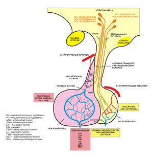
Neurosecretory cells, which are found mainly in the nucleus supraopticus and nucleus paraventricularis, send unmyelienized axons to the neurohypophysis. This connection together forms the hypothalamohypophysary tract (tractus hypothalamohypophysialis). Axons produce large dilatations filled with basophilic neurosecretory granules, which are called Herring bodies. The contents of the granules are synthesized in neurosecretory neurons and released into the vicinity of the capillaries in the neurohypophysis. Granules store oxytocin, which is produced in the nucleus paraventricularis and antidiuretic hormone (ADH, vasopressin) from the nucleus supraopticus. Pituicytes are glial cells that fall between macroglia that surround unmylienized axons, providing them with support.
Functions of the Posterior pituitary gland hormones
- Oxytocin: Uterine contractions during labour and stimulates contraction of the myoepithelial cells to drain milk into the lactiferous duct during lactation.
- Antidiuretic hormone: Regulates the bodies electrolyte balance, blood pressure and kidney functioning by increasing the reabsorption of water at the distal convoluted tubules of the kidneys.
Blood supply[edit | edit source]
The anterior lobe and posterior lobe have the same venous drainage (anterior and posterior hypophyseal veins), but have separate arterial blood supply.
Blood comes to the pituitary gland through aa. hypophysiales superiores et inferiores, branches of a. carotis interna and drains through the pituitary portal veins. To the adenohypophysis comes a. hypophysialis superior, which in front of it branches around the eminentia mediana hypothalami in the primary capillary plexus. It is then collected in the pituitary portal vein, which leads the blood to the secondary capillary plexus running through the adenohypophysis. This venous blood is collected into the vv. hypophysiales, which flows into the sinus cavernosus and through it into the v. jugularis interna. This phenomenon, when there is an insertion of the capillary plexus between the vessels of the same type, is called the rete mirabile, in this case it is the rete mirabile venosum (the plexus is inserted between the veins).
This arrangement serves to the fact that hormones affecting the activity of the pituitary gland, released by neurosecretory neurons of the hypothalamus into the primary capillary plexus, reach the place of action - the adenohypophysis - through the blood.
To the neurohypophysis comes a. hypophysialis inferior, which also goes to the vv. hypophysiales.
Activity regulation[edit | edit source]
Both stimulation and inhibition of the release of hormones from the pituitary gland are controlled by the hypothalamus, which communicates with the gland via neurotransmitters. These neurotransmitters are secreted into the hypophyseal portal vessels. This allows the hypothalamic hormones to remain concentrated, and not be diluted in the systemic circulation.
Stimulating hormones include:
- corticoliberin - corticotropin-releasing hormone (CRH, corticotropine-releasing hormone);
- gonadoliberin - gonadotropin-releasing hormone (GnRH, gonadotropine-releasing hormone);
- thyreoliberin - thyreotropin-releasing hormone (TRH, thyrotropine-releasing hormone).
Inhibitory hormones:
- somatostatin inhibits somatotropin output;
- dopamine inhibits the release of prolactin.
Links[edit | edit source]
Related articles[edit | edit source]
Literature used[edit | edit source]
- JUNQUEIRA, L. Carlos and Chosé CARNEIRO. Basics of histology. 7th edition. Jinočany : H&H, 1999. ISBN 8085787377.
- GRIM, Miloš and Rastislav DRUGA. Basics of anatomy. 1st edition. Praha : Galén, 2005. ISBN 8072623028.
- MARTÍNEK, Jindřich and Zdeněk VACEK. Histological atlas. 1st edition. Praha : Grada, 2009. ISBN 9788024723938.
External links[edit | edit source]
- Pituitary Gland, University of Pittsburgh, Department of Neurological Surgery
- The Pituitary Gland - Structure - Vasculature - TeachMeAnatomy


