Brain stem
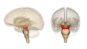
The brain stem connects rostrally to the spinal cord (medulla spinalis) and consists of the medulla oblongata, Varoli's bridge (pons Varoli) and the midbrain (mesencephalon). The brain stem has a number of structures in common, and a number of pathways or nuclei run through the entire stem and do not respect its division into the medulla oblongata, pons and midbrain at all.
Medulla oblongata[edit | edit source]
The elongated spinal cord (medulla oblongata) is a continuation of the spinal cord in the rostral direction and, as its most caudal part, already belongs to the brain. The dividing line between the spinal cord and the spinal cord is the decussatio pyramidum. The border of the medulla oblongata and Varola's bridge is the sulcus bulbopontinus, a groove running horizontally at the rostral end of the medulla oblongata. Ventrally, the elongated spinal cord is arched into two parallel, longitudinal mounds - pyramides medullae oblongatae, which contain the white matter of the pyramidal pathway, i.e. tractus corticospinalis. The median anterior fissure runs between them.
Laterally, there are paired elevations, called olives, on the elongated spinal cord. Dorsal to the olive lie the pedunculi cerebellares inferiores. The pedunculi cerebellares are generally thick bundles of white matter through which the pathways connect the stem (in this case the medulla oblongata) to the cerebellum. The peduncles diverge into a V-shape, and between them is spread the velum medullare inferius, which is a thin plate, an outgrowth of the ependyma. The free end of the vela is joined by the tela choroidea ventriculi quartii, a fibrous plate containing the choroid plexus, which forms the cerebrospinal fluid, here to IV. cerebral ventricles.
Tela choroidea ventriculi IV. it is not whole, it contains several holes. They are apertura median ventriculi quarti (foramen Magendi) and paired aperturae laterales ventriculi quartii (foramina Luschkae). These openings form a communication between the ventricular system and the subarachnoid space of the brain and allow the circulation of cerebrospinal fluid. The dorsal surface of the elongated spinal cord is also arched into two bumps - tuberculum gracile and tuberculum cuneatum. They contain the nuclei of the same name, which are the final station of the spinal fasciculus gracilis and cuneatus, which lead the main sensitive pathways of the brain. The pathways connect here and continue further into the higher levels of the brain. The canalis centralis runs through the oblongata, which opens cranially into the IV. cerebral ventricles. The following cranial nerves depart from the medulla oblongata – IX, X, XI and XII.[1]
Pons Varoli[edit | edit source]
Varol's bridge is a continuation of the medulla oblongata rostral to the sulcus bulbopontinus. It forms an oval arch on the ventral side of the brainstem. It passes cranially into the midbrain. The ventral side of the bridge is smooth and convex. The sulcus basilaris, formed by the course of the artery of the same name, leads through the medium. Laterally, the bridge passes freely in the pedunculi cerebellares medii, analogous to the pedunculi cerebellares inferiores of the medulla oblongata. Like the medulla oblongata, the pons is also related to the cranial nerves, which have their nuclei here. Nerves V, VI, VII and VIII depart from the bridge.
Mesencephalon[edit | edit source]
The middle brain (mesencephalon) is the most rostral part of the brainstem, it connects to Varol's bridge.
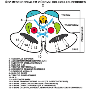
It connects the rhombencephalon to the diencephalon. It measures about 2cm in length. Practically all of it is covered by the hemispheres of the hindbrain, only its ventral part is visible as the so-called crura cerebri (partes anteriores pedunculi cerebri) – massive trunks containing white matter. The aquaeductus mesencephali (Sylvii) runs through the middle brain – a narrow channel, carrying the cerebrospinal fluid, after the distance from IV. chambers.
Aquaeductus m. is lined with a layer of gray matter substantia grisea centralis.
The midbrain can be divided into several parts - tectum mesencephali and pedunculus cerebri (which consists of tegmentum mesencephali and crura cerebri).
The pedunculus cerebri is ventral to the aquaeductus m.
Crura cerebri (partes anteriores pedunculi cerebri) - also part of pedunculus cerebri. They are lateral and sink under the optic tract into the base of the brain.
Between the two trunks is the fossa interpeduncularis. Its surface is perforated by the course of a series of vessels, therefore it is called substantia perforanta posterior (interpeduncularis). Be careful not to confuse the substantia perforanta anterior, which is part of the hindbrain!
In the fossa interpeduncularis, medially from the pedunculi, the third oculomotor nerve emerges.
Ventral to the fossa interpeduncularis are the corpora mamillaria, which already belong to the diencephalon. These cranial nerves depart from the midbrain – III, IV (II runs here already as a pathway, not the optic nerve). The cranial ends of the reticular formation also extend here.
The tectum lies dorsal to the aquaeductus mesencephali. It contains two pairs of bumps - colliculi superiores et inferiores.
They are involved in the visual and auditory pathways and continue as the brachium colliculi superioris et inferioris to the corpus geniculatum laterale and mediale diencephala.
Caudal to the colliculi extend the pedunculi cerbellares superiores, another in the series of cerebellar tracts. As with the pedunculi cerebellares inferiores, the velum, more precisely, the velum medullare superius, forming the cranial part of the IV ceiling, is opened here. chambers. Cranial to the tectum is the area pretectalis, which already belongs to the diencephalon.
Nuclei of the tectum mesencephali:
- predominantly sensitive pathways (sight and hearing)
a) Nc. colliculi superioris - fibers from the retina of the eye as well as motor and somatosensitive
b) Nc. colliculi inferioris - auditory cortex center
c) Nc. posterior commissures
d) Ncc. pretectales - nc. optic tract
Tegmentum (partes posteriores pedunculi cerebri) – ventral to tecta, part of pedunculus cerebri.
The boundary between the crura and the tegmentum is formed by the substantia nigra.
Substantia nigra - pigment in the perikarya, the outer part goes to the crura and has a net-like appearance (pars reticularis) and the inner part goes to the tegmentum (pars compacta) produces dopamine.
It contains a number of important pathways and nuclei. On the side, a gentle elevation is visible - the trigonum lemnisci, where the lemniscus medialis runs.
Nuclei of the tegmentum mesencephali:
- mainly motor paths
a) Nc. ruber - oval, large, reddish, between substantia nigra and aquaeductus, regulation of limb movements
b) Nc. nervi oculomotorii - next to the aquaeductus, a set of several nuclei, sends somatomotor fibers to the oculomotor nerve, which innervate 4 of the 6 oculomotor muscles, controls the movements of the eyeball
c) Nc. accessorius n. oculomotorii - dorsal from nc. nervi oculomotorii, visceromotor parasympathetic fibers, ciliaris muscle and sphincter pupillae, movements of the eyeball
d) Nc. interstitialis Cajal - small, dorsal from nc. nervi oculomotorii, related to the gray matter of the substantia grisea centralis and to the rostral part of the formatio reticularis, part of the fasciculus longitudinalis medialis
e) Nc. Darkshevics - small fibers from the cerebellum, cortex of the telencephalon, vestibular nuclei of the rhombencephalon
f) Nc. nervi trochlearis - small, next to the substantia grisea centralis, caudal from n. III, somatomotor fibers innervate the obliquus superior bulbi muscle
g) Nc. mesencephalicus nervi trigemini - long and slender, it goes here from the rhombencephalus, laterally from the aquaeductus, sensitive muscle and joint receptors of the masticatory muscles, artic. temporomandibularis and oculomotor muscles
Pathways of the mesencephalon:
- afferent to ncc. collicules superiores from the retina of the eye, from the spinal cord, from the rhombencephalon, from the visual cortex
- to colliculi inferioris from ncc. cochlear
Links[edit | edit source]
External links[edit | edit source]
- Mefanet - Atlas Mozkový kmen a mozeček
- DUBOVÝ, Petr. Základy neuroanatomie a nervových drah : Multimediální podpora výuky klinických a zdravotnických oborů [online]. Portál Lékařské fakulty Masarykovy univerzity [online], ©2007. Poslední revize 3.6.2011, [cit. 2011-11-27]. ISSN 1801-6103. <http://portal.med.muni.cz/clanek-442-zaklady-neuroanatomie-a-nervovych-drah.html>.
Related articles[edit | edit source]
References[edit | edit source]
- ČIHÁK, Radomír. Anatomie III. 2., upr. a dopl vydání. Praha Grada Publishing, spol. s. r. o.. 2004. 673 s. ISBN 80-247-1132-X.


