Anal fistula
An anal fistula is a pathological connection between the skin of the perineum and the rectum or anal canal. It occurs 8 times more often in men than in women. The total prevalence is about 7/100,000 people. The average age of an affected individual is 40 years.
Etiology[edit | edit source]
In the area of the rectum and perineum, there are also fistulas that may not be related to the rectum (they could be related to the female genitalia, urethra, or prostate). All these fistulas can be classified as congenital and acquired.
- Congenital fistulas result from a defect in rectal development.
- Acquired fistulas:
- – 80–90% arise as a result of perianal abscesses caused by infections of the anal glands (so-called cryptoglandular theory).
- – 10–20% have a different etiology: dermoid cyst, perineal trauma, Crohn's disease, pelvic inflammation, TBC, carcinomas, radiation therapy, actinomycosis, chlamydial infections, or other sexually transmitted diseases.
- Rare fistulas: horseshoe-shaped fistulas that surround the anal canal. Most often they are transsphincteric. If unrecognized, they often relapse.
Divisions[edit | edit source]
Fistulas can be classified according to their relationship to the rectum - rectal and periproctal (perianal). Furthermore, we divide them into complete and incomplete according to whether they have external and internal orifices (complete) or only one (incomplete, more often external). A complex (complicated) fistula is a fistula that has an inner orifice that extends above the puborectalis muscle or its tract surrounds more than 3/4 of the external sphincter.
According to the anatomical position of the tract, fistulas can be classified as:
- Intrasphincteric (subcutaneous, submucosal)
- Intersphincteric
- Transsphincteric
- Extrasphincteric
According to Parks: intrasphincteric, transsphincteric, suprasphincteric, extrasphincteric
Symptoms[edit | edit source]
Symptoms include secretions, itching, and wetting of the fistula's surroundings as well as contamination of underwear with stool or pus. Pain occurs when the contents stagnate and the drainage stops. Swelling, bleeding, and feelings of fullness and pressure in the rectum may also occur. Subfebrile symptoms may also occur. It is common for affected individuals to experience symptomatic periods interrupted by asymptomatic periods of various lengths of time. An internal fistula may not manifest clinically at all.
Diagnostics[edit | edit source]
A thorough anamnesis with a focus on ano-rectitis and abscesses in this area prior to the examination is important. The search for symptoms of Crohn's disease can also help diagnose anal fistulas. Physical examination is of the greatest importance:
- Inspection – the external orifice of the fistula is found. Fistulas may have multiple external orifices but usually have only one internal orifice. The distance of the orifice from the anus indicates the type of fistula. When it is closer to the anus, the fistula is most likely subcutaneous, whereas if it is further away from the anus, it is more likely to be a complicated fistula.
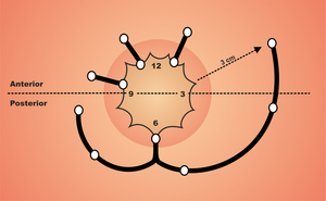
- Goodsall's rule – if a transverse line across the anus is observed and the fistula's external orifice is posterior to this line, then the internal orifice is in the midline posteriorly with a horse-shoe track (at No. 6). Fistulas that have an external orifice anterior to the line have a straight path and open radially to approximately the same position. However, Goodsall's rule does not always apply, especially in women.
- Palpation: one can feel induration in the surroundings of the fistula. During rectal examinations, one can sometimes feel the internal orifice.
- Anoscopy, proctoscopy, and colonoscopy allow for the evaluation of the rectal mucosa, the localization of the internal orifice of the fistula, and the exclusion of tumors or inflammatory bowel disease.
- Fistula probing: one can use a stick probe to determine the course of the fistula in a non-violent manner. If the localization of the inner orifice is unclear, one can use a solution of hydrogen peroxide, betadine, or methylene blue.
- Fistulography: a contrast agent is used for fistulas in Crohn's disease, recurrence, or complicated course. It reveals the fistula's branching and course. However, the yield of the examination is low
- US: endosonographic examination has recently been widely used, replacing CT examination, which is of little use for diagnosis of fistulas. US imaging can be used for primary diagnosis and perioperatively.
- NMR: nuclear magnetic resonance is the most accurate method for the imaging complicated fistulas, especially in Crohn's disease.
Treatment[edit | edit source]
Conservative techniques:
- In the past, conservative therapy involving the application of sclerosing agents (e.g., AgNO3), was widely used, but it had a poor outcome, so it is no longer used.
- Today, various fibrin tissue adhesives are used. The most effective adhesives are combined with ATBs.
Surgical techniques:
- Fistulotomy, fistulectomy: These similar procedures are performed on simple low fistulas, which do not have a complicated course. After drainage of the fistula and internal orifice, a groove probe is inserted into the fistula and the fistula is dissected. The surrounding tissue is excised and the base is excochleated, and the tissue sample can be taken for analysis. The wounds then undergo marsupialization or are left to heal via secondary healing. These techniques are associated with large tissue damage and a higher percentage of postoperative incontinence. Therefore, there are various modifications of operations to preserve the integrity of the external sphincter (Parks fistulectomy).
- Seton technique – (Hippocratic elastic ligature, cutting seton) is a technique used in higher transsphincteric, extra- and suprasphincteric fistulas. After the identification of the external and internal orifices, a non-absorbable, elastic fiber (silicone, rubber) is threaded through the fistula and knotted under a slight pull. The fiber gradually cuts through the sphincter, and it must be tightened several times. The healing process requires 6-8 weeks and fibrotic tissue gradually forms behind the cut fiber. The technique can also be performed subcutaneously. Indications must be considered. There is a greater risk of incontinence in ventral fistulas in women.
- Seton drainage is used for complex, multiple fistulas, and fistulas in Crohn's disease. This technique is similar, but the fiber is fistula introduced loosely without tension. Inelastic materials can also be used. Thanks to seton drainage, the fistula gradually matures, followed by its excision. In Crohn's disease, this technique is used for a long-term (weeks to months) drainage of fistulas with abscesses.
- Mucosal sliding flap (advancement flap) has the best results and can be used with almost all types of fistulas. It is particularly effective at treating complicated fistulas. Healing is preferably per primam. It does not cause anal deformities. The method involves a fistulectomy outside the sphincters, an excision of the internal orifice, which is followed by the construction of a sliding mucosal tube, which is pulled into the defect and fastened with sutures around the circumference. The external defect is left to secondary healing.
- Anal fistula plug: the use of special biological anal plugs that allow the fistula to heal can also be performed.
New operating techniques:
- LIFT – Ligation of the Intersphincteric Fistula Tract: a new, relatively simple technique of intersphincteric ligation.
- VAAFT – Video Assisted Anal Fistula Treatment: an endoscopic examination of the fistula tract (fistuloscopy) with visualization of branching and possibility of internal orifice treatment using a stapler or cutter.
- Fistula cauterization using RFA or laser.
References[edit | edit source]
Related Articles[edit | edit source]
Sources[edit | edit source]
- HORÁK, Ladislav. Praktická proktologie. 1. edition. Praha : Grada, 2013. ISBN 978-80-247-3595-5.
- REJCHRT, Stanislav. Přístup k paciento s píštělemi gastrointestinálního traktu. Folia Gastroenterologica et Hepatologica [online]. 2007, y. 5, vol. 5, no. 1, p. 19-29, Available from <http://www.pro-folia.org/files/1/2007/1/rejchrt.pdf>.
- BARTOŠKA, Petr. Perianální píštělě [online]. ZdravíE15, ©2007. [cit. 2014-04-01]. <https://web.archive.org/web/20160331222721/http://zdravi.e15.cz/clanek/postgradualni-medicina/perianalni-pistele-319038>.


