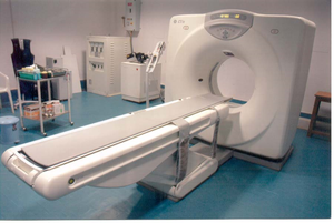
Computed tomography
Computed Tomography[edit | edit source]
Computed tomography (CT scan, CAT scan, sometimes incorrectly computer tomography) is a medical imaging method allowing us to produce a series of virtual cross-sectional images of the whole human body with the use of X-rays. The final image is a result of tomographic reconstruction of a sequence of X-ray projections, obtained from different angles. CT scan can be used to view hard tissues, as well as soft tissues, e.g. spleen, pancreas, kidneys, brain, muscles. Using the computed tomography imaging method we are only able to detect such pathological processes, which differ in density from their surroundings, whether with or without the administration of a contrast agent.
Process[edit | edit source]
By computed tomography we are producing virtual transverse "slices" of a laying patient. The patient is fixated on a mobile bed (the patient must remain stationary on the bed), which is passing through a stand with a scanner. On one side of the scanner there is a source of X-rays (X-ray tube), and on the opposite side is a set of scintillation counters (detectors). Most modern CT scans are equipped with scintillation counters that form a complete stationary vertical ring around the patient.
The patient is being screened in one level (plane) spot after spot. The X-ray tube is pulsating, one pulse lasts 1-4 ms. X-rays are passing through the patient, where they are being partially absorbed. In a given position of the patient an exposition is made. Then the information about the cut-down of the X-rays obtained by the detectors is saved in the computer. After that the whole system of X-ray tube and detectors rotates by a certain angle and the whole process starts again. After the completion of all the scanning cycles, the data from all of the detectors are saved into the computer. These data are then processed by the computer and the final tomogram is made based on the values of the absorption coefficients from individual spots of the tissues of the given "slice".
There are two modalities of computed tomography: fan-beam and cone-beam. In the case of fan-beam CT the X-ray tube as well as the detectors are spun around the area being scanned, whereas in the cone-beam tomograph only the X-ray tube is spun and the detectors are located on the perimeter of the machine.
Classification of computed tomography based upon the mechanical motion required to collect data:
1. generation - single X-ray source producing pencil beam translated across patient, X-rays are detected by one detector, which rotates along with the X-ray tube
2. generation - X-ray source emits radiation over a fan-shaped angle, after the translation across patient it is measured by a number of detectors arrayed in a line, significantly faster than the first generation
3. generation - X-ray tube emits radiation in a wide fan-shaped beam (similar to 2. generation), but the translated beam is detected by a large number of scintillation detectors arrayed in an arc-shape in several rows, scanning of several "slices" at the same time - "multi-slice CT", used most frequently nowadays in modern medicine
4. generation - stationary ring of detectors around patient, rotating X-ray tube
5. generation - coronary-CT angiography, X-rays - electron beam
History[edit | edit source]
The fundamental influence on the invention of CT scan had Wilhelm Conrad Röntgen, who discovered X-rays in 1895. Nowadays we still use X-rays for RTG imaging. The inventor of the actual CT scanning is Godfrey Newbold Hounsfield. Independantly of Hounsfield´s discovery, Allan McLeod Cormack made the same invention in 1979. Both of them were awarded the Nobel prize. Previously the examination took 20 minutes, today it takes tens of seconds.
Use of CT in healthcare[edit | edit source]
In urgent medicine, there is no contraindication for the use of CT. It is widely used in diagnostics and in therapeutic operations. Using X-rays, it displays the internal organs and skeleton.
Advantages of CT[edit | edit source]
The great advantage of CT scanning is the fact, that it allows us to visualise and distinguish low-contrast soft tissues. This is due to two reasons: Scintillation detectors which detect X-rays passing through the patient´s body are vere sensitive, and the data provided by them are processed by the computer very quickly. The computer then expresses them as absorption coefficient values, which drastically increases the accuracy of the examination.
Before or during examination, contrast agents are often administered, in order to highlight the differences between normal and pathological tissue.
Sources[edit | edit source]
https://www.wikiskripta.eu/w/V%C3%BDpo%C4%8Detn%C3%AD_tomografie
- KYMPLOVÁ, Jaroslava. Katalog metod v biofyzice [online]. [cit. 2012-09-20]. <https://portal.lf1.cuni.cz/clanek-793-katalog-metod-v-biofyzice>.
- SEIDL, Zdeněk. Radiologie pro studium i praxi. Praha: Grada, 2012. ISBN 978-80-247-4108-6.
References:[edit | edit source]
- Základy výpočetní tomografie (Doc.RNDr. Roman Kubínek, CSc.)
- Výpočetní tomografie (Czech Wikipedia)

