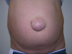
Umbilical hernia
Hernia umbilicalis is a condition where there is no complete closure in the area where the umbilical cord entered the abdominal cavity. The umbilical cord ensured the nutrition of the child during intrauterine development.
Physiological hernia occurs when the intestinal loop rapidly lengthens, especially its cranial arm. At the same time, the liver also enlarges, and the volume of the peritoneal cavity is now too small to contain the entire contents of the developing midgut. Thus, during the 6th week of embryonic development, the intestinal loop reaches the extraembryonic coelom of the umbilical cord and a physiological hernia occurs. This defect will disappear on its own after birth. It is usually until the 16th month of age. If the opening does not retract on its own, the umbilical gate (hilus) is open and the intra-abdominal organs penetrate the subcutaneous tissue through the opening. Most often, it is a loop of the small intestine and the omentum.
Occurrence[edit | edit source]
Umbilical hernia is the most frequent disease in children that requires surgery. Hernias occur twice as often in boys than in girls.
Symptoms[edit | edit source]
In the place of the navel, a painless, soft and elastic formation bulges out when the intra-abdominal pressure increases (e.g. when crying or screaming). Sometimes it is inconspicuous, so it escapes attention during preventive examinations at the pediatrician. But it is often the size of a pea or a cherry.
Clinical examination is performed both with the patient lying down and standing. A physical examination the so-called 4 (5) P – look (aspect), touch (palpation), tap (percussion) and listen (auscultation). For determining the size and location is an essential aspect. Percussion with auscultation is important in recognizing hernia complications (peritoneal irritation, intestinal obstruction, etc.). Palpation is used to specify the size of the hernia, to clarify the hernia gate, to determine whether it is a replaceable or irreplaceable hernia, helps to estimate the organs in the hernia, and can provide much more information.
Paraclinical examination are only useful for a small number of hernias. Their overuse is financially demanding and can be harmful due to a number of factors. The local finding is exceptionally necessary to specify USG or CT examination - Spiegel's hernia in an obese patient. If intestinal obstruction or "perforation peritonitis" related to hernia complications is suspected, a native image of the abdomen (if possible, in a standing position) is appropriate → however, it is not necessary if the hernia is clearly narrowed. For large and massive hernias, it is necessary to perform a thorough cardiopulmonary examination, including spirometry and other special methods.
Treatment[edit | edit source]
Treatment is operative if this defect does not correct itself by the 16th month of the child's age. The most favorable period for surgery is around the 2nd-5th. year of the child's age. If operated on in adulthood, the operation is more difficult. Only newborns can do without surgery, in which patch fixation can be performed, but it is uncomfortable, or a so-called hernia belt is attached, which has a Velcro fastener.
Complications[edit | edit source]
Incarceration is the most dangerous complication that can occur with a hernia. It is the entrapment of the visceral organ in the hernia sac in such a way that the vascular supply is damaged. The low-pressure venous system is first oppressed and edema, hemostasis to passive hemorrhagic infarction occurs. Furthermore, the high-pressure arterial system is compressed with subsequent ischemic necrosis. A transudate begins to be found in the hernia sac, which becomes infectious and spreads to the surrounding area. Intestinal perforation may occur. The inflammation can penetrate either into the abdomen and cause peritonitis or into the abdominal wall and lead to its phlegmon or abscess.
Links[edit | edit source]
Related articles[edit | edit source]
Used literature[edit | edit source]
- SADLER, Thomas W. Langmanova lékařská embryologie. 10. edition. Praha : Grada Publishing, a.s., 2011. 414 pp. pp. 250. ISBN 978-80-247-2640-3.
- PÁČ, Libor – HORÁČKOVÁ, Ladislava. Anatomie pohybového systému člověka. 1. edition. Brno : Masarykova univerzita, 2009. pp. 146. ISBN 978-80-210-4953-6.
Sources[edit | edit source]
- AMED. Pupeční kýla - hernia umbilicalis. Amed - dětstká chirurgie Praha [online]. Praha [cit. 2019-01-12]. Dostupné z: http://www.chirurgie-amed.cz/kyla/pupecni-kyla
- NOVÁK, Karel. Pupeční a ventrální kýly [online]. Nové Město, 2001, Available from <http://www.cls.cz/dokumenty2/os/r030.rtf>.

