Types of joint injuries and principles of treatment
From WikiLectures
Distortion (sprain)[edit | edit source]
Distortion (sprain) occurs through indirect action or direct violence when the physiological range of motion in a given joint is exceeded.
Clinical picture[edit | edit source]
- partial rupture or distension of the capsule or ligaments. It can also be hemarthroses
- the joint remains stable
- soreness, swelling, restriction of movement, hematoma
Therapy[edit | edit source]
- puncture a larger effusion, evacuate the hepatoma and flush the cavity with cold saline or mesocaine (in case of severe pain)
- immobilisation, according to disability, lighten the joint, give NOA, ice
- we can indicate arthroscopy for the knee
Subluxation[edit | edit source]
A subluxation is an incomplete dislocation. It is caused by more violence than distortion. The bones are in a so-called subluxation position, when the joint surfaces only partially touch.
Clinical picture[edit | edit source]
- Injury to the capsule and ligaments is greater than with distortion.
- The joint is slightly unstable, but spontaneous reduction often occurs.
Therapy[edit | edit source]
- Rigid immobilisation is necessary for a period of 3–6 weeks.
- Relief, icing, NOA
- In more difficult cases – operative revision with ligament suture.
Luxation[edit | edit source]
Luxation (dislocation) occurs in case of significant force on the joint (possibly less force in case of predisposition), a serious disorder of congruence occurs . Reduction can be spontaneous, but usually the joint is dislocated.
According to the mechanism of formation, we distinguish sprains:
- Traumatic – caused by sudden and strong violence that breaks the stabilizing fibrous structures of the joint.
- Habitual – arises as a result of primary or secondary functional disorders or anatomical structure of the joint.
- Pathological – in case of long-term changes in the joint (damage of the joint surfaces during paralysis, loosening of the joint capsule during chronic inflammation).
- Congenital - basis in the presence of congenital dysplasia (hip).
Clinical picture[edit | edit source]
- swelling, hematoma, significant pain
- we monitor innervation, blood supply to the periphery, momentum
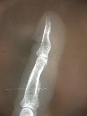
- we are investigating whether it is a dislocation fracture on the X-ray
Therapy[edit | edit source]
- perform under local or general anesthesia
- after repositioning, we check stability and detect damage to soft tissues
- immobilize the joint
- subsequent rehabilitation is important
Links[edit | edit source]
Related articles[edit | edit source]
- Injury
- Regional anesthesia, blockades
- [[General anesthesia]
Reference[edit | edit source]
- BENEŠ, Jiří. Studijní materiály [online]. [cit. 2009]. <http://jirben.wz.cz>.


