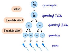
Gametogenesis procedure
 Gametogenesis is the formation of gametes - gamet (from the Greek γαμετης = married). Gametes are produced by reduction division - meiosis. The latter is evolutionarily younger than mitosis. As a result of meiosis, the gametes have a haploid set of chromosomes. They develop from the cells of the germinal epithelium of the gonads of males - testes' and of females - ovaria.
Gametogenesis takes place in two stages:
- phase of growth' (proliferation) - primordial germ cells - gametogonia - having a diploid set of chromosomes arise by mitosis;
- maturation phase - in the gonads, gametogonia produce gametes by meiosis.
The development of male and female sex cells is quite different.
Spermatogenesis[edit | edit source]
Spermatogenesis takes place during the entire period of sexual activity in two successive processes - "spermatocytogenesis" and the subsequent "spermatohistogenesis". After completion of spermiohistogenesis, spermatozoa are released into the lumen of the ducts and are passively entrained to the epididymis. Each germ cell produces four full-fledged sperm.
Spermatocytogenesis[edit | edit source]
Male sex cells - sperm - develop in the seminiferous tubules of the testiste. Their maturation begins under the influence of sex hormones during puberty. Diploid germ cells - spermatogonia - grow repeatedly, are enriched with nutrients and divide mitotically either into still undifferentiated spermatogonia A or into already differentiated spermatogonia B, which gradually mature into spermatocytes of the first order in the so-called growth stage. When the cell enters the maturation stage, the first meiotic division occurs and spermatocytes II are formed. order (prespermatids). This is followed by a brief interkinesis in which DNA replication does not occur. The second meiotic division produces spermatids – small cells with haploid sets of chromosomes. Prespermatids and spermatids remain connected by intercellular cytoplasmic bridges, ensuring synchronization of development and exchange of gene products between cells. Spermatids remain in the folds of Sertoli cells, which supply them with important substances and energy for their development.
Summary:
spermatogonia → spermatocyte of the first order → spermatocyte of the second order. order → early spermatid → late spermatid → spermatozoon
Spermatohistogenesis (spermiogenesis)[edit | edit source]
Through the final maturation of the male germ cells, referred to as spermiohistogenesis, the spermatids acquire the shape and function necessary to penetrate the egg and fertilize it:
- Golgi phase:
- small PAS positive granules accumulate in the Golgi complex and merge into the acrosomal lysosome;
- flagellum axoneme begins to form;
- nucleus chromosomes condense very tightly, DNA is fixed into a crystalloid form by basic proteins - protamines (they replace histones); all gene activity is suppressed.
- cap phase:
- acrosomal sac grows and pulls the cap-like front edge of the nucleus.
- acrosomal phase:
- a final acrosome containing hydrolytic enzymes is formed (hyaluronidase – during fertilization, it disrupts corona radiata, acid phosphatase, protease acrosin and others);
- cell rotates to point its anterior pole toward the edge of the seminiferous tubule;
- the activity of the microtubule cuff stretches the nucleus;
- distal from the centrioles grows and forms the basal body of the flagellum (the second, proximal, will form the dividing spindle in the egg after fertilization);
- mitochondria move to the proximal part of the flagellum, which they thicken to form the middle segment of the sperm;
- graduation phase:
- remaining cytoplasm discarded and phagocytosed by Sertoli cells;
- residual bodies remain from the bridges that connected the individual cells formed from one spermatogonia during development.
Oogenesis[edit | edit source]
Female sex cells - eggs' - develop in the ovaries. The human egg has a diameter of 0.1 mm, the size is species specific. The ovum develops from cells of the germ line in the cortex of the ovary. Around 2 million germ cells - oogonia - are established in the ovary. The egg, like every gamete, contains half the number of chromosomes (22 somatic + 1 sex).
The multiplication of oogonia by mitotic division begins at the end of the 2nd month and ends in the 5th month of intrauterine development in the first stage of reproductive division (meiosis). The cells of the coelom epithelium are attached to the oogonia in one layer, so-called "primordial follicles" are formed. In about 50% of oogonia, this layer is not formed, the cells die by apoptosis. The oogonia give rise to ``oocytes of the first order by mitosis (in women, their formation - the growth stage - ends as early as the 3rd month after birth). Oocytes of the first order enter the meioses. The prophase of the first meiotic division proceeds to the diplotene stage, in which the oocytes remain until the hormonal initiation of further maturation, the so-called dictyotene stage
The maturation of individual follicles then continues after puberty under the influence of hormones''. The stimulus for the continuation of the first maturation division is in some species progesterone, in others the change in the level of estrogens and progesterone during the cycle. The maturation stage takes place throughout the entire generation period (with a female in cycles of Ø 28 days – one egg matures each time). During a woman's lifetime, 300-400 eggs are released from the ovary, but only about 400-500 eggs mature. The first meiotic division (in metaphase) gives rise to two haploid cells from the developing oocyte: one oocyte II. order and one rudimentary cell – the so-called polocyte (polar body). The cell remains undivided.
The second meiotic division is completed after ovulation and after the penetration of the sperm into the egg, when from the oocyte II. order, one egg and the other polar body will be formed. At the same time, the 1st poleocyte still goes through mitosis, it divides into two, and all 3 pole bodies soon disappear and are resorbed. During the maturation of the egg before ovulation, some genes of the egg are intensively expressed. The synthesis of RNA, proteins and various storage substances takes place, some mRNA are transported to the cytoplasm, where they are stored in an inactive form and activated only after fertilization of the egg. Other storage substances, proteins and RNA of all types are transported to the egg from the cells of the follicle. Some storage substances are synthesized in the mother's liver and transported by blood to the egg. The structure of the egg and the amount of substances are thus significantly influenced by the genes of the maternal somatic cells. In the case of hormonal regulation disorders, the proliferative phase of the cycle is prolonged, referred to as "overripening" of the egg. The result is the disintegration of the cell structure and the disruption of the dividing spindle and the emergence of non-disjunctions of chromosomes and aneuploidy in the foetus.
Links[edit | edit source]
Related Articles[edit | edit source]
Source[edit | edit source]
- ŠTEFÁNEK, Jiří. Medicína, nemoci, studium na 1. LF UK [online]. [cit. 11.02.2010]. <https://www.stefajir.cz/>.


